


Deficits in strength of the hip external rotators (ER) affect trunk, hip, and knee movement patterns, potentially contributing to injury in athletes.
ObjectivesTo provide normative data on isometric torque for hip ER in athletes of three distinct sports and to determine if isometric torque for the hip ER and torque asymmetry between legs differ among sports and between sexes.
MethodsBasketball, soccer, and volleyball athletes (n=451) were evaluated. Hip ER torque was quantified bilaterally with athletes in prone and 90° of knee flexion using a hand-held dynamometer.
ResultsData are expressed as mean and 95% confidence interval. Hip ER torque values in Nm/kg for the dominant and non-dominant limbs were, respectively, 0.46 (0.44, 0.48) and 0.42 (0.40, 0.44) for male soccer athletes; 0.35 (0.32, 0.37) and 0.27 (0.25, 0.29) for male basketball athletes; and 0.37 (0.34, 0.39) and 0.35 (0.32, 0.37) for male volleyball athletes. Hip ER torque in Nm/kg for the female volleyball athletes was 0.29 (0.26, 0.33) for the dominant and 0.29 (0.25, 0.32) for the non-dominant limb. The Limb Symmetry Index for male soccer, basketball, and volleyball players was, respectively, 94% (91, 97), 81% (75, 87), and 95% (91, 99). For female volleyball players the Limb Asymmetry Index was 102% (95, 108). Male volleyball athletes showed higher torque values than female volleyball athletes.
ConclusionsThis study reported normative values for hip ER isometric torque of youth athletes. Clinicians can use the reported data as reference to identify torque deficits in athletes of the three reported sports.
Volleyball, basketball, and soccer athletes have high incidence of lower limb injuries,1,2 mostly related to jumping, landing, and cutting movements.3–5 Safe performance of these specific movements during sports practice and competitions relies on the athletes’ capacity to stabilize the hip and, consequently, minimize movement impairments of more distal joints. For example, weakness of the hip external rotators (ERs) leads to excessive femoral internal rotation during functional tasks (e.g., single-leg squat, landing) and more distal movement impairments, such as dynamic knee valgus and excessive foot pronation.6,7
Lower limb movement impairments related to hip ER weakness include knee valgus and excessive foot pronation, which are associated with patellofemoral pain syndrome8,9 and anterior cruciate ligament (ACL) injuries.3,10,11 Importantly, a study that investigated the contribution of a number of muscles responsible for core stability showed that hip ER strength was the only useful predictor of injury occurrence in intercollegiate basketball and track athletes.10
Consistent with these findings, researchers have suggested that a strengthening program targeting the hip ERs is an effective intervention to decrease knee pain and address poor lower limb kinematics during functional activities that have been related to injury risk in women.9,12,13 To effectively identify strength deficits in hip ER, to prescribe strength training as part of prevention and rehabilitation programs, and make decisions about return to sport, it is helpful to know what the expected values of hip ER strength are. Some level of muscle strength asymmetry is expected between dominant and non-dominant lower limbs, and documenting asymmetry index in uninjured athletes is crucial given the role of strength asymmetries to sports injury.14–16
The primary objective of this study was to provide normative data on hip ER torque for dominant and non-dominant legs of athletes of three different sports (volleyball, basketball, and soccer). Our secondary objectives were to determine if (1) hip ER torque and torque asymmetry between legs significantly differ among sports; and (2) between males and females.
MethodsParticipantsThis cross-sectional study was part of a larger project involving preseason assessment and lower limb injury risk profile identification of 4 different sport-clubs from the city of Belo Horizonte, Brazil and was approved by the Universidade Federal de Minas Gerais Research Ethics Committee (report number 0493.0.203.000-09). All volleyball, basketball, and soccer youth, competitive level athletes participating in the project were recruited and signed a consent form. All included athletes were regularly practicing sports and did not have pain or history of surgery to the lower limbs and trunk in the past 6 months. Failure to properly follow test instructions and/or having pain during the testing session were exclusion criteria. For those athletes under 18 years-old, the parents signed the consent form after the athlete's assent.
ProceduresBefore the test, each athlete jogged for 5-minutes for warm-up. Two trained examiners (physical therapists with 5 years of clinical practice) followed a standardized procedure to assess hip ER isometric torque of all participants.14 For the assessment, participants were lying in prone, with arms positioned along the body on the treatment table. The knee was positioned at 90° of flexion and hip at 0° (neutral) rotation; this position was monitored by an inclinometer (Starrett®) positioned below the tibial tuberosity. Both legs were randomly tested for each participant.
The examiner positioned the hand-held dynamometer (Microfet2®) using a rigid strap to eliminate measurement bias (examiner force application during the test). The dynamometer's strap was anchored to a metal rod placed perpendicular to the table (Fig. 1). Another stabilization strap was fixed around the pelvic region of the athlete to avoid pelvic movement. The dynamometer was positioned five centimeters proximal to the medial malleolus and the rigid strap limited hip external rotation range of motion (ROM).

We allowed the athletes to produce one practice isometric contraction by pulling against the rigid bar to which the dynamometer was attached with progressively increasing strength, for familiarization purposes. Subsequently, the athlete completed the test following the examiner's standard verbal command to contract the hip ER, gradually increasing strength, until the command to stop the test after 5 seconds of contraction.17 The examiner closely observed the athlete's lower limb during force production. If compensations were observed before the 5-second mark, the test was interrupted and repeated after a 15-second interval. Compensations included pelvic elevation and/or rotation, trunk rotation, extension and/or adduction of the tested lower limb and isometric flexion of the hip of the opposite lower limb.
Participants performed three isometric contractions with each leg, with 30 second interval in between repetitions. The mean value of the 3 repetitions for each leg was multiplied by the respective lever arm to obtain the magnitude of hip ER isometric torque. The lever arm was measured with a ruler from the dynamometer that was positioned 5 cm proximal to the medial malleolus, to the femur medial condyle. Torque was normalized by body mass (Nm/kg). To describe the expected asymmetry between dominant (D) and non-dominant (ND) limbs, a symmetry index was computed according to the following formula: (ND/D)*100.18 An index of 100%, indicates perfect symmetry. Indices <100 and >100 indicate stronger dominant and non-dominant leg, respectively.18 Lower limb dominance was defined through the following question: ‘‘Which leg would you use to kick a ball as hard as possible?’’
A pilot study with 6 participants (4 women and 2 men, healthy university students, with a mean age of 21.6 years, mean body mass of 62.2 kg, and mean height of 1.64 m) was conducted to assess intra- and inter-examiner reliability of the torque assessment procedure. The results showed excellent reliability (intraclass correlation coefficient intra-examiner = 0.98 and inter-examiner = 0.90), a standard error of measurement (SEM) of 0.021 Nm/kg, and a minimal detectable difference (MDD) of 0.05 Nm/kg.
Statistical analysisAll torque data are reported in Nm per unit of body weight, thus, considering the height (limb length) of the individuals and adjusted for weight. To achieve the primary objective of the study, box plots were used to provide a visual representation of the computed descriptive data (mean and 95% Confidence Interval (CI)) of body-mass normalized hip ER isometric torque (stratified by limb) and Limb Symmetry Index.
Linear mixed effect models were estimated to test hypothesis related to the secondary objectives of this study. This approach allows for the inclusion of random effects to capture and control for individual differences across participants not accounted for by fixed effects. Additionally, linear mixed effect models are robust to imbalances in group sizes. Results, however, should be interpreted as ANOVA results.
Two models were estimated. Model 1, computed with data from male athletes, tested for significant differences in hip ER torque between sports, between dominant and non-dominant limbs, and for significant sports modality × leg dominance interaction effect. Model 2, computed with data from volleyball athletes, tested for differences in hip ER torque between males and females and for significant sex × leg dominance interaction effect. Finally, simple Analyses of Variance were performed with data from male and volleyball athletes, respectively, to determine if torque asymmetry differed (a) among sports (Model 3), and (b) between males and females (Model 4). Appropriate follow-up pair-wise comparisons with Tukey correction were conducted, as needed, on the least square means estimated by each of the models.
ResultsFour hundred and fifty-one competitive level youth athletes participated in this study: 208 volleyball athletes (137 males and 71 females), 77 basketball athletes (all male), and 166 soccer athletes (all male). The mean age (in years) of male volleyball, basketball, and soccer players was, respectively, 18.3 (95% CI: 17.6, 19.1), 17.0 (95% CI: 16.2, 17.8), and 18.6 (95% CI: 18.0, 19.2). The mean age of female volleyball players was 16.4 (95% CI: 15.8, 17.0). The age range within all groups is very small and the maximal difference between sports was less than 2 years. Thus, any differences in ER torque comparisons between groups are not likely meaningfully affected by age.
Normative data: hip external rotator torque & asymmetry indexFig. 2 illustrates the typical distributions of (body-mass normalized) hip ER isometric torque (stratified by limb) and between-legs asymmetry index. Fig. 2A and B present data of male participants stratified by sports, and Fig. 2C and D present data of volleyball athletes stratified by sex. Distributions of hip ER torque scores (Fig. 2A and C) look quite symmetric for males of all three sports. The distribution for females looks right skewed, suggesting a higher prevalence of lower torque values than would be expected from a strictly normal distribution. Skewness and kurtosis scores for both dominant and non-dominant legs were, however, within acceptable ranges (<2) for all distributions. The distribution of asymmetry indices (Fig. 2B and D) looks right skewed for basketball athletes and symmetric for soccer and volleyball athletes. Again, the skewness and kurtosis scores (<2) did not suggest excessive deviations from normal in any of the distributions. Extreme outliers for torque and asymmetry index (i.e., values that were more than 3 times the interquartile range above the mean) were identified and excluded from the dataset prior to further analysis. In total, we excluded one observation of torque obtained from the dominant leg and one obtained from the non-dominant leg. We have also excluded 7 observations of Limb Symmetry Index.
 hip external rotator isometric torque for dominant and non-dominant legs (A and C) and for between limb torque symmetry index (B and D). A and B: data from male athletes in the study sample stratified by sport. C and D: data from volleyball athletes in the study sample stratified by sex. (*) indicates extreme outliers that were removed from data set.")
Distributions of (body-mass normalized) hip external rotator isometric torque for dominant and non-dominant legs (A and C) and for between limb torque symmetry index (B and D). A and B: data from male athletes in the study sample stratified by sport. C and D: data from volleyball athletes in the study sample stratified by sex. (*) indicates extreme outliers that were removed from data set.
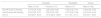
Table 1 provides the normative data for (a) hip ER torque for dominant and non-dominant limbs and (b) Limb Symmetry Index, stratified by sports and sex. Notably, intervals obtained for male athletes are smaller than 0.05 Nm/kg (the MDD of the torque variable) and only slightly greater than that for female athletes. This suggests that the sample size was sufficient to provide estimates of hip ER torque with small margin of errors, which is critical if these estimates are to be useful to guide decision making of sport physical therapists and trainers.
Normative data for body-mass normalized hip external rotator torque and symmetry index (SI).
Data are presented as mean (95% confidence interval). Hip ER torque D, hip external rotator torque for dominant leg; Hip ER torque ND, hip external rotator torque for non-dominant leg; LSI, Limb Symmetry Index (non-dominant/dominant).
Model 1 (male data) showed that hip ER torque significantly differed among male athletes of different sports, p < 0.001, and between dominant and non-dominant legs, p < 0.001. The model also returned a significant sports × leg dominance interaction effect, p < 0.001.
Pair-wise comparisons of least squares means were performed to follow-up on the interaction effect. Results showed that soccer athletes produced significantly higher torques than both volleyball and basketball athletes regardless of limb dominance, all p values < 0.0001. There was no significant difference between volleyball and basketball players regardless of limb. Differences observed between dominant and non-dominant limbs depended on sports and were only statistically significant for basketball (p < 0.01) and soccer athletes (p < 0.01).
Model 2 (volleyball data) returned a significant effect of sex, p = 0.001, with male volleyball athletes showing higher torque values than female volleyball athletes. There was no significant effect of dominance, p = 0.11, nor sex × leg dominance interaction effect, p = 0.08.
Sports and sex-related differences: limb symmetry indexModel 3 (male data) showed that Limb Symmetry Index differed statistically among sports. Pair-wise comparisons of least squares means returned significant differences in Limb Symmetry Index between basketball athletes and both volleyball and soccer athletes (p-values < 0.01), with basketball athletes showing higher asymmetry. No significant difference was observed between soccer and volleyball athletes.
Model 4 (volleyball data) returned a significant effect of sex, p = 0.005, with greater asymmetry observed for males than females.
DiscussionThis study presents normative values of hip ER torque for volleyball, basketball, and soccer athletes. The reported torque values were higher than those reported in previous studies for non-athlete population.19 These data can be used to support the identification of deficits in hip ER torque that needs to be addressed in injury prevention and rehabilitation programs specifically designed for athletes.
The normative data for hip ER torque obtained in this study provide sport-specific guidelines to identify athletes who might be in need for intervention. The results from Boling et al.19 showed that individuals with patellofemoral pain had weakness in eccentric and concentric torque compared to the asymptomatic group.19 Therefore, athletes who have values of hip ER torque outside the expected range might benefit from strength training intervention.
Our study indicates that the expected magnitudes of torque and between-limb symmetry differ across the evaluated sports, suggesting the need for sport-specific normative data. The mean torque presented by soccer athletes was higher than basketball and volleyball athletes for both legs. Interestingly, this trend might not be specific to hip ER. Bittencourt et al.15 also demonstrated that hip abductor torque was smaller in basketball athletes than indoor soccer athletes.15 Possibly, this difference is associated with the specificity of each sport. Soccer training, such as ball passing using the foot and lower limb agility work, or weight lifting training, may be more intense and focused on strengthening the lower limb when compared to training of athletes from the other two sports.
The results also indicated that differences between dominant and non-dominant leg depended on sports. In particular, significant differences between legs were not observed in volleyball athletes. Some differences in muscle torque between legs are, however, expected in basketball and soccer, with the non-dominant leg being in general less strong.
Muscle torque asymmetry between legs was greatest (lower limb asymmetry between legs) for basketball athletes and may be related to specific demands of the sport. A common task performed by basketball athletes during practice is the "lay-up". During this task, the athlete drives towards the basketball hoop handling the ball and then jumps forward (toward the hoop) using one leg and releases the ball.20 Additionally, during jumps the athlete normally has physical contact in the air with other athletes, which can lead to single-leg landing. Most typically, the athlete uses his/her dominant leg to land on, which would justify greater strength on this side. Further research is required to examine the hypothesis that strength differences between sports are related to training and performance specificities.
The effect of sex on muscle strength has been highlighted in the literature. For example, Barber-Westin et al.21 found differences between males and females for knee extensors and flexors strength. Bittencourt et al.,15 also found difference between sexes for the hip abductors in youth volleyball athletes. Sex difference could be expected due to hormonal differences during growth.22 Consistent with the literature the present study showed a significant sex-related difference in hip ER torque, female volleyball players presented lower torque values than male and males had greater asymmetry then females. Thus, prevention programs should take into account sex difference for hip ER when planning tailored exercises.
Most recent studies have used hand-held dynamometers for assessment of muscular strength, likely because of its cost-effectiveness compared to isokinetic dynamometers.14,23 Similar to the present study, Thorborg et al.23 reported excellent reliability for testing of hip ER in prone (Intraclass Correlation Coefficient (ICC) = 0.98) and recommended the use of hand-held dynamometers in clinical practice to detect hip weakness in different populations. Notably, the measures obtained with this device are static and, thus, when information about dynamic muscular strength is required, isokinetic dynamometry remains the gold standard.15 Importantly, Bazzet-Jones et al.24 noted that measurements obtained using such devices are smaller in magnitude than those obtained with an isokinetic device. After considerations of validity and reliability issues, clinicians and researchers can choose the equipment that best fits their context. However, they must use normative data obtained with a similar method to identify athletes in need for strength training. Interestingly, there is a strong correlation between hip ER isometric torque and hip extensors isometric torque (r=0.80, p<0.0001) in healthy adults and older adults, indicating that 63.8% of the total hip ER torque variance was explained by the hip extensors torque.25 Therefore, the normative data that we presented can be used to guide hip ER and extensors strength training, especially considering that gluteus maximus is the primary contributor to both hip external rotation and extension.
The findings should be considered with some limitations. We analyzed sex influence in hip ER torque only in volleyball athletes, thus future studies are needed to identify if sex differences are present in other sports. Hip ER torque was assessed using a hand-held dynamometer, which is reliable, easy to use, and inexpensive. However, our reliability study was conducted on a sample of shorter athletes. The examiners, however, were well trained and did not experience difficulty during the assessment of the study sample. Finally, it should be noted that we focused on youth athletes, who are frequently overlooked in the literature. Data should not be generalized to adult athletes, who can be the focus of future research.
ConclusionThe present study reported normative values for hip ER torque of youth athletes. Clinicians can use the reported data as reference to identify torque deficits in athletes during pre-season assessments for injury prevention purposes or during rehabilitation of sports injuries. Data also provide information on expected between-leg asymmetries in torque. A large-scale prospective study should be developed to affirm preventive actions guided by these normative data.
Conflicts of interestThe authors do not have any conflict of interest that could inappropriately influence this research.
The authors thank the following sports club for providing the athletes who participated in this study: Minas Tenis Clube, Cruzeiro-SADA, Clube Atlético Mineiro and América Futebol Clube.


