Journal Information
Vol. 26. Issue 2.
(01 March 2022)
Share
Share
Download PDF
More article options
Visits
2404
Vol. 26. Issue 2.
(01 March 2022)
Original Research
Full text access
International physical therapists consensus on clinical descriptors for diagnosing rotator cuff related shoulder pain: A Delphi study
Visits
2404
Néstor Requejo-Salinasa,b,w, Jeremy Lewisc,d,e, Lori A Michenerf, Roy La Touchea,b,g, Rubén Fernández-Matíash,i,
Corresponding author
ruben.fernanmat@gmail.com

Corresponding author at: Postal Address: Street Cruz de Guadalajara 4, 2°A, 28805 Alcalá de Henares, Madrid, Spain.
, Juan Tercero-Lucasj, Paula Rezende Camargok, Marcus Batemanl, Filip Struyfm, Jean-Sébastien Royn,o, Anju Jaggip, Timothy Uhlq, Leanne Bissetr, Craig A. Wassingers, Robert Donatellit, Melina Nevoeiro Haiku, Enrique Lluch-Girbésj,v
a Department of Physical Therapy, Superior Center for University Studies La Salle, Autonomous University of Madrid, Madrid, Spain
b Motion in Brains Research Group, Institute of Neuroscience and Movement Sciences (INCIMOV), Superior Center for University Studies La Salle, Autonomous University of Madrid, Madrid, Spain
c School of Health and Social Work, University of Hertfordshire, Hertfordshire, United Kingdom
d Therapy Department, Central London Community Healthcare National Health Service Trust, London, United Kingdom
e Department of Physical Therapy & Rehabilitation Science, College of Health Sciences, Qatar University, Doha, Qatar
f Division of Biokinesiology and Physical Therapy, University of Southern California, Los Angeles, United States
g Neuroscience and Craniofacial Pain Institute (INDCRAN), Madrid, Spain
h Research Unit, Hospital Universitario Fundación Alcorcón, Alcorcón, Madrid, Spain
i Research Institute of Physical Therapy and Pain, University of Alcala, Alcala de Henares, Madrid, Spain
j Department of Physical Therapy, University of Valencia, Valencia, Spain
k Laboratory of Analysis and Intervention of the Shoulder Complex, Department of Physical Therapy, Universidade Federal de São Carlos, São Carlos, SP, Brazil
l Derby Shoulder Unit, Orthopaedic Outpatient Department, Royal Derby Hospital, Derby, United Kingdom
m Department Rehabilitation Sciences and Physiotherapy, Faculty of Medicine and Health Sciences, University of Antwerp, Antwerp, Belgium
n Department of Rehabilitation, Faculty of Medicine, Université Laval, Quebec City, Quebec, Canada
o Center for Interdisciplinary Research in Rehabilitation and Social Integration, Quebec Rehabilitation Institute, Quebec City, Quebec, Canada
p Royal National Orthopaedic Hospital NHS Trust, London Borough of Harrow, London, United Kingdom
q Department of Physical Therapy, University of Kentucky, Lexington, United States
r School of Allied Health Sciences, Griffith University, South East Queensland, Australia
s Department of Physical Therapy, East Tennessee State University, Johnson City, Tennessee, United States
t Modern Athletic Science, Las Vegas, United States
u Department of Physical Therapy, Center of Health and Sport Science, Universidade do Estado de Santa Catarina, Florianópolis, Santa Catarina, Brazil
v Pain in Motion Research Group, University of Valencia, Valencia, Spain
w International Doctoral School, Rey Juan Carlos University, Alcorcón, Spain
Ver más
Highlights

  • 18 clinical descriptors for diagnosing RCRSP were defined across six domains.

  • The results summarize the current knowledge about diagnosis of RCRSP.

  • The results could be useful to standardize the diagnosis of RCRSP.

This item has received
Article information
Abstract
Full Text
Bibliography
Download PDF
Statistics
Figures (3)
Show moreShow less
Additional material (1)
Abstract
Background

There is a lack of standardized criteria for diagnosing rotator cuff related shoulder pain (RCRSP).

Objective

To identify the most relevant clinical descriptors for diagnosing RCRSP.

Methods

A Delphi study was conducted through use of an international physical therapists expert panel. A 3-round Delphi survey involving an international panel of physical therapists experts with extensive clinical, teaching, and research experience was conducted. A search query was performed in Web of Science, along with a manual search, to find the experts. The first round was composed of items obtained from a previous pilot Delphi study along with new items proposed by the experts. Participants were asked to rate items across six clinical domains using a five-point Likert scale. An Aiken's Validity Index ≥ 0.7 was considered indicative of group consensus.

Results

Fifteen experts participated in the Delphi survey. After the three rounds, consensus was reached on 18 clinical descriptors: 10 items were included in the “subjective examination” domain, 1 item was included in the “patient-reported outcome measures” domain, 3 items in the “diagnostic examination” domain, 2 items in the “physical examination” domain”, and 2 items in the “functional tests” domain. No items reached consensus within the “special tests” domain. The reproduction of symptoms in relation to the application of load, the performance of overhead activities, and the need of active and resisted movement assessment were some of the results with greatest consensus.

Conclusion

In this Delphi study, a total of 18 clinical descriptors across six clinical domains were agreed upon for diagnosing RCRSP.

Keywords:
Assessment
Delphi study
Diagnosis
RCRSP
Rotator cuff
Shoulder pain
Full Text
Introduction

Shoulder pain is the third leading cause of musculoskeletal pain in primary care consultations.1–3 Incidence rates range from 14.7 to 29.3 per 1000 person-years2–4 with a lifetime prevalence that can reach up to 70% in the general population.5 Shoulder pain is common among people between 45 and 55 years old4 and, despite heterogeneity in the terms used to identify specific pathologies,6,7 the most frequent finding among patients with shoulder problems is pain originating from the rotator cuff and other subacromial structures.8,9

Historically, the term used to describe this condition was subacromial impingement syndrome.10 Findings from recent surgical research11 have challenged the basis for the acromion as the cause of the pathology leading to pain. In addition, research has shown that exercise therapy has the same short, medium, and long-term benefit as acromioplasty in combination with exercise therapy,12 further questioning impingement as the primary mechanism of symptoms.13 A series of clinical terms have emerged to actively move away from the flawed term impingement syndrome. These terms include, among others, subacromial pain syndrome,14,15 rotator cuff disease,16 and rotator cuff-related shoulder pain (RCRSP),13 that were introduced to move away from an incorrect pathoanatomical explanation for the presenting symptoms. Particularly, RCRSP was suggested as an overarching term that includes subacromial pain syndrome, subacromial impingement syndrome, rotator cuff tendinopathy, and subacromial bursitis.13 Non-traumatic RCRSP includes non-traumatic partial and full-thickness tears of the rotator cuff and traumatic RCRSP involves traumatic tears of these structures.13 This term was proposed aiming to reduce patients’ concerns related to aberrant acromial spurs causing symptoms and thus beliefs that surgery was needed for symptoms resolution. In this regard, there is recent evidence showing that diagnostic labels for shoulder pain may influence people's perceived need for surgery,17 as well as how patients can perceive the potential effectiveness of physical therapy.18 In particular, Zadro et al.17 found that the labels “rotator cuff tear” and “subacromial impingement syndrome” were those that most encouraged people to consider surgery. Interestingly, there was no significant difference in terms of considering surgery between these labels and the label “RCRSP”, so it is not clear that the use of this term solves the apparent problem.

The term RCRSP acknowledges that we currently don't know the underlying cause of the shoulder pain. It is one of many painful shoulder presentations which is characterized by pain and/or weakness most commonly experienced in shoulder elevation and external rotation, absence of referred pain (i.e. from cervical spine), and occurrence related with a change in shoulder loading behavior and potential changes in lifestyle.13,19 It is important to note that to establish a diagnosis of RCRSP, differential diagnosis with other clinical conditions such as shoulder instability or frozen shoulder syndrome should be made as their clinical presentation may sometimes be similar.20,21

Establishing a functional diagnosis is the primary objective within the professional practice of physical therapists22; this is often challenging in people with shoulder pain. The coexistence of multiple pathologies,23 the lack of reliability of orthopedic tests,24–27 the lack of direct association between imaging findings and clinical symptoms,9,28–30 as well as the possible implication of proximal segments31 make it very difficult to pinpoint the exact source of shoulder pain.

Regarding RCRSP diagnosis, no standardized diagnostic criteria are currently available.32 Some proposals for diagnosing RCRSP have been published,13,15,33,34 but in no study a strong enough methodology was used enabling the proposed diagnostic criteria to be accepted as universal for this clinical condition. This fact, added to the need to accurately differentiate RCRSP from other shoulder clinical conditions, justified the aim of this study which was therefore to identify the most relevant clinical descriptors for RCRSP based on the opinion of an international panel of experts with a high level of clinical, teaching, and research experience.

Methods

A three-round online Delphi survey was employed to obtain a consensus on which clinical descriptors are necessary and sufficient for RCRSP diagnosis. Clinical descriptors are defined as findings obtained throughout the examination and assessment processes that may guide the diagnosis of a clinical condition.35,36 Initially developed by Dalkey,37 the Delphi method is commonly used in health science research as a reliable way to reach a consensus on clinical issues.38,39 The Delphi method involves both a workgroup and a participant (respondent) group of experts.40 The study was approved by the Centro Superior de Estudios Universitarios La Salle Ethics Committee (ES) (CSEULS-PI-025/2020). The research was conducted according to the Declaration of Helsinki.

Participants

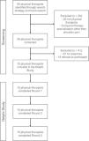
The expert panel comprised physical therapists purposely selected based on their experience and knowledge in RCRSP.41,42 Only physical therapists were included in the study to make this study more relevant to physical therapy teaching, research, and practice. Additionally, trying to establish an acceptable diagnosis/label such as RCRSP may help patients to understand their problem and facilitate adherence to a treatment (i.e., exercise)18 that has been shown to be as effective as a surgical procedure.43 To assemble a representative group of experts, a list of inclusion criteria was established and the Web Of Science database was queried using a search of relevant terms related to the study topic (Supplementary File - Table 1). Additionally, a manual search was performed to verify that a series of additional experts proposed by the working group also met the inclusion criteria. The selection process is depicted in Fig. 1.

Fig. 1.

Flow diagram of experts.

(0.38MB).
RCRSP, Rotator Cuff Related Shoulder Pain; ROM, range of motion

The working group consisted of five investigators, physical therapists (NRS, RFM, ELG, RLT, JTL), who were responsible for designing the survey and collecting and analysing the data from each round of questions.

Procedure

For all three Delphi rounds, the experts received an invitation by email with a link to an online questionnaire. The experts had 3 weeks to complete each round, with reminders emailed weekly.

First, the expert panel was sent three documents via email: a letter inviting them to participate in the study, an informed consent document created by the working group, and a questionnaire. The first part of this questionnaire included a list of sociodemographic questions. The second part was composed of items structured into six domains (subjective examination, patient-reported outcome measures [PROMs]), diagnostic examination, physical examination, special tests, and functional tests) as described in a previous DELPHI study.36 These initial items were chosen after two members of the working group (ELG and JTL) carried out a three-round non-published pilot Delphi study (with the same methodology as stated below for the final Delphi study) with 10 shoulder experts from February to March 2019 (Table 1). The use of a previous pilot study has been reported in some DELPHI studies44,45 and it has been suggested that it could ensure greater rigor, especially regarding the design of the first round questions.46 For the first round of the current Delphi study, participants were asked to rate the items from the pilot study using a five-point Likert scale (Strongly Disagree, Disagree, Neutral, Agree, Strongly Agree). In addition, in this first round, experts were invited to add items to each of the six examination domains.

Table 1.

Clinical descriptors obtained in the previous pilot Delphi study.

Subjective examination 
The onset may be insidious or traumatic. 
Pain with movement of the arm – typically abduction or overhead activities is indicative of RCRSP. 
Deltoid region pain is indicative of RCRSP. 
Age typically over 40 years. 
May follow a period of increased activity. 
Patient-reported outcome measures 
Not relevant to the diagnosis. 
Diagnostic examination 
Diagnostic image is not indicated unless history of cancer or trauma. 
The management (surgical and non-surgical) of RCRSP is not influenced by the image. 
Physical examination 
Active ROM assessment of the shoulder should be done in patients suspected of RCRSP. 
Muscle strength tests should be done in patients suspected of RCRSP. 
Pain on resisted abduction is indicative of RCRSP. 
Pain often on resisted external rotation is indicative of RCRSP. 
Patients with RCRSP often have limited internal rotation but not always. 
Patients with RCRSP should not have pain or limited range of movement in cervical extension-rotation. 
The absence of findings on palpation of the cervical spine is indicative of RCRSP. 
Full range of passive external rotation is indicative of RCRSP. 
Full range of motion of the cervical spine is indicative of RCRSP. 
Special test 
Any active test resisted (or not) that reproduces pain in a systematic way (same conditions: directionality, activity, load, speed, position, functional activity). Not necessary to use any "special" test. It's no more useful to use orthopedic tests looking for other conditions (e.g., lag sign, instability, …). 

RCRSP, rotator cuff related shoulder pain; ROM, range of motion.

The level of agreement among the experts was analysed by means of the Aiken's V coefficient of validity (V). This coefficient is used to quantify the content validity or relevance of an item with respect to a content domain evaluated by several experts’ judgement. Aiken's V is calculated as the ratio of the sum of agreement score obtained from all authors for a given item, with respect to the maximum possible score (i.e. maximum value of the Likert scale * number of experts rating that item). The value of Aiken's V ranges from 0 to 1, the latter representing perfect agreement. An Aiken's V ≥ 0.7 was considered reflective of group consensus, as recommended for Delphi studies.47

The second round of questions included items that reached at least a 0.7 validity index in the first round and the suggestions made by the expert panel members. No open-ended questions were included, and the experts were asked to indicate their degree of agreement with each proposed item using the previously mentioned five-point Likert scale.

For round 3, participants were informed of the results from round 2 using descriptive statistics and asked to reconsider their degree of agreement with each item before expressing their final opinion. Each participant was, therefore, asked to re-grade the criteria that reached consensus in round 2 using the same Likert-type scale.

Data analysis

All analyses were performed with statistical software R version 4.1.0 (R Core Team (2021). R: A language and environment for statistical computing. R Foundation for Statistical Computing, Vienna, Austria. URL https://www.R-project.org/). Mean and standard deviation (SD) and absolute and relative frequencies were used for the descriptive analyses of the continuous and nominal variables, respectively. The agreement between experts was analysed using the Aiken's Validity Index (Aiken's V), an agreement index for ordinal data with scores ranging from 0 (no agreement) to 1 (perfect agreement).48

Results

After three consecutive rounds, conducted between March and May 2020, a consensus was reached among experts across the six initially proposed examination domains.

Expert panel results

For the first round, 56 physical therapists who were experts in the study topic were selected using the inclusion criteria, and 29 participants responded to the initial email. Of the 29 respondents, 14 declined to participate in the study for different reasons (most common: not having enough time) and 15 (26.78%) completed the three rounds of questions. The final expert panel consisted of 15 physical therapists from diverse work fields (research, clinical practice, education, or mixed). The group had an average ± standard deviation of 23.3 ± 8.1 years of experience with RCRSP and an average of 24.9 ± 17.3 scientific publications related to shoulder pain. Demographic information for the expert panel members is provided in Table 2.

Table 2.

Characteristics of the Delphi participants.

Sex (male:female)  7:8 
Age* (years)  48.5 ± 9.9 
Clinical experience managing patients with RCRSP (years)*  23.3 ± 8.1 
Patients with shoulder pain treated per month*  48.5 ± 79.1 
Patients with RCRSP treated per month*  25.9 ± 40.8 
Country   
Australia 
UK 
Canada 
USA 
Brazil 
Belgium 
Professional area   
Clinical practice 
Research 
Education 
Research and education 
Research and clinical practice 
Research, clinical practice, and education  10 
Highest academic degree   
Bachelor 
Master 
PhD  12 
Current workplace   
Public health system 
Private clinic 
University 
Public health system and private clinic 
University and private clinic 
Public health system and pro bono 
University outpatient clinic 

*Mean ± standard deviation or frequency; RCRSP, Rotator Cuff Related Shoulder Pain; pro bono, services to those who are unable to afford them.

Delphi survey results

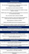
At the end of the three rounds, a total of 24 RCRSP-related items were obtained (Supplementary File -Table 2). They were distributed amongst the six diagnostic domains as follows: subjective examination (n = 11), PROMs (n = 1), diagnostic examination (n = 4), physical examination (n = 6), and functional tests (n = 2). No items reached consensus within the “special tests” diagnostic domain. Two items from the "subjective examination" were merged (“Pain with movement of the arm – typically abduction or overhead activities is indicative of RCRSP” and “Pain and weakness with elevation or overhead activities are indicative of RCRSP“) and one from "diagnostic examination" (“Diagnosis imaging is required if the patient has a history of trauma”) was eliminated after reaching consensus among the working group because it was highly similar to another item. A total of 22 clinical descriptors for diagnosing RCRSP were obtained. However, some were unified and grammatically edited by the working team to simplify understanding, leaving a total of 18 items (Fig. 2). The original items of Round 3 without unification or editing are presented in Supplementary File - Table 2.

Fig. 2.

Final descriptors of the Delphi study.

(0.72MB).

Overall, 44.4% (8/18) of the items from the initial Delphi pilot study remained at the end of the third round and 92.31% (24/26) of the items that reached consensus in round 2 remained in round 3. The complete item selection process is represented in Fig. 3.

Fig. 3.

Flow diagram of the Delphi study.

(0.42MB).
Subjective examination

In round 1, 20% (1/5) of the items from the initial pilot study did not reach the Aiken's V required for consensus (Supplementary File - Table 3). The experts proposed 18 additional items (Supplementary File - Table 4). In round 2, 10 items were discarded and a total of 12 items (54.6%) were kept for round 3 (Supplementary File - Table 5), where 91.6% (11/12) reached consensus (Supplementary File - Table 2).

Patient-reported outcomes measures

The one item from the pilot study did not reach the necessary degree of consensus (Supplementary File - Table 3) and seven new items were proposed by the respondent group (Supplementary File - Table 4). In the second round, only one item (14.3%) reached consensus (Supplementary File - Table 5), which was also maintained at the end of round 3 (Supplementary File - Table 2).

Diagnostic examination

In round 1, 50% of the items from the pilot study (1/2) did not reach sufficient consensus (Supplementary File - Table 3) and experts proposed 12 additional items (Supplementary File - Table 4). In the second round, 30.7% of the items (4/13) reached consensus (Supplementary File - Table 5); these four items maintained the consensus among the experts at the end of round 3 (Supplementary File - Table 2).

Physical examination

Of the nine initial items proposed, five (44.4%) did not reach consensus in round 1 (Supplementary File - Table 3). Ten more items were proposed by the experts (Supplementary File - Table 4). From round 2, 42.9% (6/14) of the items continued to round 3 (Supplementary File - Table 5) and remained at the end of the study (Supplementary File - Table 2).

Special tests

The single initially proposed item did not reach the necessary level of consensus (Supplementary File - Table 3). Ten items were proposed by experts in this first round (Supplementary File - Table 4). In round 2, only one item (10%) reached consensus (Supplementary File - Table 5) but failed to do so at the end of the third round (Supplementary File - Table 2).

Functional tests

No items were included from the pilot study in this diagnostic domain. However, the experts proposed nine items in round 1 (Supplementary File - Table 4) of which two (22.2%) earned Aiken's V scores sufficient for consensus in round 2 (Supplementary File - Table 5). These same two items remained at the end of round 3 (Supplementary File - Table 2).

Discussion

The objective of this study was to obtain a consensus on the most relevant diagnostic criteria for RCRSP. A total of 18 clinical descriptors across six domains were identified using the Delphi method. This approach is proven valid for obtaining expert opinions on a given topic and is widely used in health science research.40,42,49 The Delphi method has three important features: anonymity, controlled feedback, and statistical group response.37,50

Respondent group characteristics

The respondent group included 15 experts. There is currently no consensus on the ideal sample for a panel of experts with some authors recommending a minimum of 15,51 10,52 or even seven members.53 The quality of the panel of experts seems to be more important than the number when judging the level of representativeness of the results obtained in a Delphi study.49,54 Additionally, the criteria for defining an expert within a Delphi study are not clearly established.55 Levels of knowledge and professional experience have been proposed as main criteria.55 After analysing the characteristics of the panel members, their years of clinical experience treating people with RCRSP (23.3 ± 8.1), the number of scientific publications in a topic related to shoulder pain (24.9 ± 17.3), and the percentage of experts with PhD as the highest professional degree (80%), the quality of the panel of experts of this Delphi study seems high. This fact is important when considering the results.

Subjective examination

The subjective examination is such a fundamental part of a physical therapist assessment that 75 to 83% of diagnostic decisions are reached based on its results.56,57

Pain related to mechanical load emerged as one of the main descriptors indicative of RCRSP in the subjective examination domain. Although the origin of RCRSP is multifactorial,13 poor load management (e.g., excessive loading, alterations to regular loading on subacromial structures) seems to be the most determining causal factor58 and plays a critical role in its progression.59 Furthermore, although the nociceptive sources of RCRSP are not only tendinous,13,23 it seems likely that the pain behavior in RCRSP is similar to that described for tendinopathies (e.g., on/off pain behavior dependent on the amount of load applied).60 A clear evidence of the influence of mechanical load on the genesis of RCRSP is that this condition is related to the performance of activities involving load in shoulder elevation,61–63 and is more prevalent on the dominant side, in active, working populations61 and overhead sports.64,65 In addition, RCRSP is more prevalent in individuals who have performed overhead activities for a long time.66–69

Regarding the location of the pain, the experts agreed that the most frequent area reported by those with RCRSP is the deltoid region, which is in line with a previous consensus report34 and experimental pain models.70 Pain reaching the forearm has also been reported in people with RCRSP.71 To the authors’ knowledge, the utility of pain distribution (e.g., using pain drawings) to assist with the differential diagnosis of shoulder pain has not fully been investigated. As experimental pain models have considerable limitations as models for clinical shoulder pain,72 future research may focus on exploring differences between patients in terms of clinical pain distribution.

Another item obtained by consensus was that RCRSP affects sleep. The relationship between the presence of shoulder pathology and sleep disturbances has been already established in other studies.73,74 Overall, aspects related to a patient's lifestyle such as sleep quality can be relevant in people with musculoskeletal pain.75,76 For example, according to the majority of the studies, sleep deprivation produces hyperalgesic changes.77 Furthermore, poor sleep can compromise the physiological processes underlying tendon recovery.78 Sleep disturbances are frequently associated with anxiety and depression, which highlights the value of performing a psychological evaluation in people with RCRSP.79,80 Also, there seems to be a dose and time-dependent relationship between tobacco consumption and the appearance of pathological changes in the rotator cuff,81 shoulder pain, and rotator cuff tear size.82 Other lifestyle-related factors (e.g., body mass index, alcohol consumption, and comorbidities like diabetes) may also be related to the development of RCRSP.66,83–85

The most frequent age of RCRSP onset related to tendon changes is between 45 and 55 years old.4 As such, being older than 50 years is a significant factor for developing rotator cuff tendinopathy among active populations.66 Moreover, being over 60 years old is a risk factor for rotator cuff tears.86 However, the item “age typically over 40″ did not reach consensus in the last round of questions (Supplementary file - Table 2). Rotator cuff disorders can appear in a wide age spectrum. Some authors even consider “two different rotator cuff diseases,” differentiating between patients under 40 years old with rotator cuff tendinopathy and older adults suffering from full-thickness tears.87 The main differences between these two populations are the potential for tissue healing, etiology, activity levels, physical demands and long-term expectations of recovery.87

Regarding age, the item “Could be secondary to atraumatic or microtraumatic instability in younger patients (< 40 years)” also reached consensus. It seems that in young people the development of rotator cuff tears may have a traumatic or atraumatic origin, this latter typical of overhead sports in the context of shoulder “microinstability.”87,88 The concept of shoulder microinstability is used to describe excessive laxity of the anterior capsule secondary to repeated movements of shoulder abduction and external rotation (i.e. overhead sports) which leads to abnormal glenohumeral biomechanics and internal shoulder impingement.89 The role of shoulder microinstability in the development of rotator cuff disorders is however controversial and some authors even consider that excessive laxity of the shoulder may act as a protective mechanism for avoiding impingement.90

The absence of neurological symptoms was reported by experts to be indicative of RCRSP, which agrees with a previous consensus report.34 Collecting sufficient data during the subjective examination can help to rule out a neurological component to the generation of symptoms.91

Patient-reported outcome measures (PROMs)

There are several PROMs for assessing shoulder pain and function, such as the Disabilities of the Arm, Shoulder and Hand (DASH) questionnaire, the quick DASH, the Shoulder Pain and Disability Index (SPADI), the Constant (Murley) Score (CS), and the American Shoulder and Elbow Surgeons (ASES).92,93 Some questionnaires including the Rotator Cuff-Quality Of Life and the Western Ontario Rotator Cuff (WORC) index have been developed specifically for people with RCRSP.94 In the current Delphi study, PROMs were considered useful for guiding treatment and prognosis but not to make a diagnosis of RCRSP.95 Shoulder pain questionnaires have demonstrated to be valid, reliable, and responsive to changes93,96,97 but some are not specific to the shoulder joint (i.e., DASH) and few are specific to RCRSP.94

Diagnostic imaging

Diagnostic imaging was considered necessary for people with RCRSP in three situations: red flags, history of trauma, and lack of response to a minimum of 3 months of conservative treatment. Imaging is widely used in medical practice for diagnosing patients with musculoskeletal pain.98–100 In people with RCRSP, magnetic resonance and ultrasound imaging have proven to be valid for diagnosing massive rotator cuff tears, with comparable levels of sensitivity and specificity.101,102 However, their sensitivity decreases with less extensive rotator cuff lesions.101,103,104 Furthermore, there is still controversy about the utility of imaging for guiding clinical decisions in people with shoulder pain including RCRSP, because it correlates poorly with symptoms.9,28,29,105 In patients with shoulder pain, it is difficult to identify the exact structure responsible for the symptomology due to the coexistence of multiple tissue alterations.32,106 However, radiological examination can help to rule out red flags.34 Finally, if we accept that exercise is key in the management of RCRSP107 and a minimum of 12 weeks is recommended to determine whether the results thereof are satisfactory,34,108 it follows that radiological examination be requested only after a lack of response to a minimum 3 months of exercise.

Physical examination

There was consensus amongst the experts that pain with resisted movements, in particular in shoulder abduction and external rotation, is indicative of RCRSP. This finding is described by others,13,34,109 which together suggests that pain with resisted movements is key for diagnosing RCRSP. Depending on the stage of pathology in which the patient with RCRSP is58 and the level of tissue sensitization23,110 pain could probably be reproduced with other resisted movements (i.e., resisted flexion).

Assessment of muscle strength was reported as necessary for suspected RCRSP. In fact, RCRSP has also been called "weak and painful shoulder".21,34 Subacromial pain induces alterations in the electromyographic activity of shoulder musculature and its ability to produce strength.111–114 Although deficits in shoulder abduction and external and internal rotation strength have been described in those with RCRSP,115–117 there is conflicting evidence.118

Special tests

No special tests achieved the level for consensus at round 3 required to become a relevant clinical descriptor for RCRSP diagnosis. This finding is consistent with the current literature questioning the usefulness of orthopedic tests in the diagnosis of shoulder pain.24–26 Most orthopedic tests used for diagnosing shoulder pain,24–26 including RCRSP,119–122 have low diagnostic accuracy. This is likely due to the inability to isolate and specifically stress one single anatomic structure with any special test,13 the coexistence of several altered anatomical structures,23,123 and potential changes in the mechanosensitivity of local tissues.110 Therefore, currently, it is recommended to stop teaching and using special tests to establish the origin of shoulder pain.27

Interestingly, the item "depending on the condition some tests may be needed" was very close to reaching consensus in the final round (Aiken's V = 0.68). Some special tests and test clusters have been shown, for example, to improve the clinical diagnostic process for arriving at a pathology-based diagnosis in people with shoulder pain.124 In our opinion, such tests should be a component of a bigger picture, not a standalone diagnostic tool and are likely to be more useful for detecting full-thickness rotator cuff tears.

Functional tests

Performance-based functional tests are performed by clinicians in an attempt to quantify and discriminate impairments in patient's body functions.125 Several upper-extremity functional tests have been described and evaluated in the shoulder region both at rehabilitation and injury-prevention settings126 and their psychometric properties have been recently summarized.125

Experts considered functional tests to be relevant for patients with RCRSP reporting subtle symptoms or at end-stage rehabilitation. However, no functional test in isolation reached a sufficient level of consensus to be useful for diagnosing RCRSP. Based on these results, functional tests may then be useful for assessing people with RCRSP with low levels of irritability.127 Functional testing was also considered useful by experts as a basis for symptom modification. Symptom modification procedures based in part on symptom reproduction with patient-specific functional movements have been recommended for assessing people with RCRSP.128

As a final consideration, the diagnosis of RCRSP is, to a certain extent, a diagnosis by exclusion. This implies that other conditions must be ruled out before accepting a diagnosis of RCRSP as valid. Interestingly, experts did not reach consensus, for example, on screening the cervical spine129 or assessing passive shoulder movements130 to determine the involvement of the cervical spine as a source of pain contribution or exclude a frozen shoulder, respectively. The absence of distal neurovascular symptoms is another parameter that should be taken into account when excluding other pathologies.131

Research strengths and limitations

This study involved a highly experienced panel of experts who were chosen using a systematic search strategy to avoid excessive selection bias. Furthermore, the preliminary pilot study likely strengthens the validity of the final results. On the other hand, the low response rate in the initial round (26.8%) might represent a limitation when considering the external validity of the proposed descriptors. Secondly, the decision of including only physical therapists in this study could consequently affect the external validity of the diagnostic criteria across other health professionals who manage shoulder pain (e.g. general practitioners, rheumatologists, sports doctors, orthopedic surgeons). Further studies are needed to validate the proposed clinical descriptors across other health professions.

Conclusions

This is the first Delphi study to our knowledge to establish a standardized consensus among physical therapists on diagnostic criteria for RCRSP. A total of 18 clinical descriptors across six examination domains were obtained for diagnosing RCRSP. The results of this study could be useful to standardize the diagnosis of this condition both in clinical and research settings. In addition, this would help to improve the management and outcome of patients with RCRSP. The clinical descriptors obtained here should be reviewed and updated regularly to reflect developments in diagnostically relevant technology and clinical information.

Funding

This research received no funding.

Acknowledgements

The authors would like to thank the experts for their effort in participating in the study.

References
[1]
M. Urwin, D. Symmons, T. Allison, et al.
Estimating the burden of musculoskeletal disorders in the community: the comparative prevalence of symptoms at different anatomical sites, and the relation to social deprivation.
Ann Rheum Dis, 57 (1998), pp. 649-655
[2]
D.A.W.M. Van Der Windt, B.W. Koes, B.A. De Jong, L.M. Bouter.
Shoulder disorders in general practice: incidence, patient characteristics, and management.
Ann Rheum Dis, 54 (1995), pp. 959-964
[3]
S.D.M. Bot, J.M. van der Waal, C.B. Terwee, et al.
Incidence and prevalence of complaints of the neck and upper extremity in general practice.
Ann Rheum Dis, 64 (2005), pp. 118-123
[4]
K. Greving, O. Dorrestijn, J.C. Winters, et al.
Incidence, prevalence, and consultation rates of shoulder complaints in general practice.
Scand J Rheumatol, 41 (2012), pp. 150-155
[5]
J.J. Luime, B.W. Koes, I.J.M. Hendriksen, et al.
Prevalence and incidence of shoulder pain in the general population; a systematic review.
Scand J Rheumatol, 33 (2004), pp. 73-81
[6]
J. Anderson.
Industrial rheumatology and the shoulder.
Rheumatology, 26 (1987), pp. 326-328
[7]
A. Bjelle.
Epidemiology of shoulder problems.
Baillieres Clin Rheumatol, 3 (1989), pp. 437-451
[8]
A. Cadogan, M. Laslett, W.A. Hing, P.J. Mcnair, M.H. Coates.
A prospective study of shoulder pain in primary care: prevalence of imaged pathology and response to guided diagnostic blocks.
BMC Musculoskelet Disord, 12 (2011), pp. 119
[9]
R.P.G. Barreto, J.P. Braman, P.M. Ludewig, L.P. Ribeiro, P.R. Camargo.
Bilateral magnetic resonance imaging findings in individuals with unilateral shoulder pain.
J Shoulder Elb Surg, 28 (2019), pp. 1699-1706
[10]
C.S. Neer 2nd.
Anterior acromioplasty for the chronic impingement syndrome in the shoulder: a preliminary report.
J Bone Jt Surg Am, 54 (1972), pp. 40-41
[11]
M. Paavola, A. Malmivaara, S. Taimela, et al.
Subacromial decompression versus diagnostic arthroscopy for shoulder impingement: randomised, placebo surgery controlled clinical trial.
BMJ, 362 (2018), pp. k2860
[12]
S. Ketola, J. Lehtinen, I. Arnala.
Arthroscopic decompression not recommended in the treatment of rotator cuff tendinopathy: a final review of a randomised controlled trial at a minimum follow-up of ten years.
[13]
J. Lewis.
Rotator cuff related shoulder pain: assessment, management and uncertainties.
Man Ther, 23 (2016), pp. 57-68
[14]
A. Papadonikolakis, M. Mckenna, W. Warme, B.I. Martin, F.A.M. Iii.
Published evidence relevant to the diagnosis of impingement syndrome of the shoulder.
J Bone Jt Surg Am, 93 (2011), pp. 1827-1859
[15]
R. Diercks, C. Bron, O. Dorrestijn, et al.
Guideline for diagnosis and treatment of subacromial pain syndrome.
Acta Orthop, 85 (2014), pp. 314-322
[16]
S. Whittle, R. Buchbinder.
Rotator cuff disease.
Ann Intern Med, 162 (2015), pp. ITC1-ITC14
[17]
J.R. Zadro, M. O’Keeffe, G.E. Ferreira, et al.
Diagnostic labels for rotator cuff disease can increase People’s perceived need for shoulder surgery: an online randomized controlled trial.
J Orthop Sport Phys Ther, 51 (2021), pp. 401-411
[18]
A. Cuff, C. Littlewood.
Subacromial impingement syndrome – what does this mean to and for the patient? A qualitative study.
Musculoskelet Sci Pract, 33 (2018), pp. 24-28
[19]
G. Burne, M. Mansfield, J. Gaida, J. Lewis.
Is there an association between metabolic syndrome and rotator cuff related shoulder pain? A systematic review.
BMJ Open Sport Exerc Med, 5 (2019),
[20]
P.W. McClure, L.A. Michener.
Staged approach for rehabilitation classification: shoulder disorders (STAR-Shoulder).
Phys Ther, 95 (2015), pp. 791-800
[21]
M.J. Kelley, M.A. Shaffer, J.E. Kuhn, et al.
Shoulder pain and mobility deficits: adhesive capsulitis: clinical practice guidelines linked to the international classification of functioning, disability, and health from the orthopaedic section of the american physical therapy association.
J Orthop Sports Phys Ther, 43 (2013), pp. A1-31
[22]
N.J. Zimny.
Diagnostic classification and orthopaedic physical therapy practice: what we can learn from medicine.
J Orthop Sport Phys Ther, 34 (2004), pp. 105-115
[23]
B.J.F. Dean, S.E. Gwilym, A.J. Carr.
Why does my shoulder hurt? A review of the neuroanatomical and biochemical basis of shoulder pain.
Br J Sports Med, 47 (2013), pp. 1095-1104
[24]
E.J. Hegedus, A.P. Goode, C.E. Cook, et al.
Which physical examination tests provide clinicians with the most value when examining the shoulder? Update of a systematic review with meta-analysis of individual tests.
Br J Sports Med, 46 (2012), pp. 964-978
[25]
S.Ø. Gismervik, J.O. Drogset, F. Granviken, M. Rø, G. Leivseth.
Physical examination tests of the shoulder: a systematic review and meta-analysis of diagnostic test performance.
BMC Musculoskelet Disord, 18 (2017), pp. 41
[26]
P. Salamh, J. Lewis.
It is time to put special tests for rotator cuff–related shoulder pain out to pasture.
J Orthop Sport Phys Ther, 50 (2020), pp. 222-225
[27]
P. Salamh, J. Lewis.
It is time to put special tests for rotator cuff–related shoulder pain out to pasture.
J Orthop Sport Phys Ther, 50 (2020), pp. 222-225
[28]
T.K. Gill, E.M. Shanahan, D. Allison, D. Alcorn, C.L. Hill.
Prevalence of abnormalities on shoulder MRI in symptomatic and asymptomatic older adults.
Int J Rheum Dis, 17 (2014), pp. 863-871
[29]
G. Tran, P. Cowling, T. Smith, et al.
What imaging-detected pathologies are associated with shoulder symptoms and their persistence? A systematic literature review.
Arthritis Care Res., 70 (2018), pp. 1169-1184
[30]
H. Minagawa, N. Yamamoto, H. Abe, et al.
Prevalence of symptomatic and asymptomatic rotator cuff tears in the general population: from mass-screening in one village.
[31]
D.G. Sueki, J.A. Cleland, R.S. Wainner.
A regional interdependence model of musculoskeletal dysfunction: research, mechanisms, and clinical implications.
J Man Manip Ther, 21 (2013), pp. 90-102
[32]
A.R. Watts, B. Williams, S.W. Kim, D.C. Bramwell, J. Krishnan.
Shoulder impingement syndrome: a systematic review of clinical trial participant selection criteria.
Shoulder Elb, 9 (2017), pp. 31-41
[33]
J. Hermans, J.J. Luime, D.E. Meuffels, M. Reijman, D.L. Simel, S.M.A. Bierma-Zeinstra.
Does this patient with shoulder pain have rotator cuff disease? The rational clinical examination systematic review.
JAMA - J Am Med Assoc, 310 (2013), pp. 837-847
[34]
C. Littlewood, M. Bateman, C. Connor, et al.
Physiotherapists’ recommendations for examination and treatment of rotator cuff related shoulder pain: a consensus exercise.
Physiother Pract Res, 40 (2019), pp. 87-94
[35]
E.J. Hegedus, B. Stern, M.P. Reiman, D. Tarara, A.A. Wright.
A suggested model for physical examination and conservative treatment of athletic pubalgia.
Phys Ther Sport, 14 (2013), pp. 3-16
[36]
M.P. Reiman, K. Thorborg, K. Covington, C.E. Cook, P. Hölmich.
Important clinical descriptors to include in the examination and assessment of patients with femoroacetabular impingement syndrome: an international and multi-disciplinary Delphi survey.
Knee Surgery, Sport Traumatol Arthrosc, 25 (2017), pp. 1975-1986
[37]
N. Dalkey.
An experimental study of group opinion: the Delphi method.
[38]
M. Falzarano, G. Pinto Zipp.
Seeking consensus through the use of the Delphi technique in health sciences research.
J Allied Health, 42 (2013), pp. 99-105
[39]
S. Keeney, F. Hasson, H. Mckenna.
Consulting the oracle: ten lessons from using the Delphi technique in nursing research.
J Adv Nurs, 53 (2006), pp. 205-212
[40]
F. Hasson, S. Keeney, H. McKenna.
Research guidelines for the Delphi survey technique.
[41]
S.S. McMillan, M. King, M.P. Tully.
How to use the nominal group and Delphi techniques.
Int J Clin Pharm, 38 (2016), pp. 655-662
[42]
E. Hohmann, J.C. Brand, M.J. Rossi, J.H. Lubowitz.
Expert Opinion Is Necessary: delphi Panel Methodology Facilitates a Scientific Approach to Consensus.
Arthrosc - J Arthrosc Relat Surg, 34 (2018), pp. 349-351
[43]
G. Nazari, J.C. MacDermid, D. Bryant, G.S. Athwal.
The effectiveness of surgical vs conservative interventions on pain and function in patients with shoulder impingement syndrome. A systematic review and meta-analysis.
[44]
T.B. Hassan, D.B. Barnett.
Delphi type methodology to develop consensus on the future design of EMS systems in the United Kingdom.
Emerg Med J, 19 (2002), pp. 155-159
[45]
H.L. Hung, J.W. Altschuld, Y.F. Lee.
Methodological and conceptual issues confronting a cross-country Delphi study of educational program evaluation.
Eval Program Plann, 31 (2008), pp. 191-198
[46]
N. Clibbens, S. Walters, W. Baird.
Delphi research: issues raised by a pilot study.
Nurse Res, 19 (2012), pp. 37-44
[47]
C. Okoli, S.D. Pawlowski.
The Delphi method as a research tool: an example, design considerations and applications.
Inf Manag, 42 (2004), pp. 15-29
[48]
L.R. Aiken.
Three coefficients for analyzing the reliability and validity of ratings.
Educ Psychol Meas, 45 (1985), pp. 131-142
[49]
C. Powell.
The Delphi technique: myths and realities.
J Adv Nurs, 41 (2003), pp. 376-382
[50]
N. Dalkey, O. Helmer.
An experimental application of the Delphi method to the use of experts.
Manage Sci, 9 (1963), pp. 458-467
[51]
H.A. Linstone, M. Turoff.
DelphiMethod: Techniques and Applications.
Reading: Addison-Wesley, (1975),
[52]
A.L. Delbecq, A.H. Van de Ven, D.H. Gustafson.
Group Techniques for Program Planning: A Guide to Nominal Group and Delphi Processes.
IL: Scott, Foresman and Company, (1975),
[53]
H.A. Linstone.
The Delphi technique.
Handbook of Futures Research, CT: Greenwood, (1978), pp. 271-300
[54]
K.A. Bantel.
Comprehensiveness of Strategic Planning: the Importance of Heterogeneity of a Top Team.
Psychol Rep, 73 (1993), pp. 35-49
[55]
J. Baker, K. Lovell, N. Harris.
How expert are the experts? An exploration of the concept of “expert” within Delphi panel techniques.
[56]
J.R. Hampton, M.J.G. Harrison, J.R.A. Mitchell, J.S. Prichard, C. Seymour.
Relative contributions of history-taking, physical examination, and laboratory investigation to diagnosis and management of medical outpatients.
Br Med J, 31 (1975), pp. 486-489
[57]
M.C. Peterson, W. Virginia, J.H. Holbrook, Smith De Von Hales;, N. Lee, L.V Staker.
Contributions of the history, physical examination, and laboratory investigation in making medical diagnoses.
West J Med, 156 (1992), pp. 163-165
[58]
J. Lewis, K. McCreesh, J.-.S. Roy, K. Ginn.
Rotator cuff tendinopathy: navigating the diagnosis-management conundrum.
J Orthop Sports Phys Ther, 45 (2015), pp. 923-937
[59]
T.J. Roberts, N. Konow.
How tendons buffer energy dissipation by muscle.
Exerc Sport Sci Rev, 41 (2013), pp. 186-193
[60]
E. Rio, L. Moseley, C. Purdam, et al.
The pain of tendinopathy: physiological or pathophysiological?.
Sport Med, 44 (2014), pp. 9-23
[61]
J. Bodin, C. Ha, A.P. le Manac'h, et al.
Risk factors for incidence of rotator cuff syndrome in a large working population.
Scand J Work Environ Heal, 38 (2012), pp. 436-446
[62]
J. Bodin, C. Ha, J.-.F. Chastang, et al.
Comparison of risk factors for shoulder pain and rotator cuff syndrome in the working population.
Am J Ind Med, 55 (2012), pp. 605-615
[63]
Y. Roquelaure, J. Bodin, C. Ha, et al.
Personal, biomechanical, and psychosocial risk factors for rotator cuff syndrome in a working population.
Scand J Work Environ Heal, 37 (2011), pp. 502-511
[64]
D.J. Lin, T.T. Wong, J.K. Kazam.
Shoulder injuries in the overhead-throwing athlete: epidemiology, mechanisms of injury, and imaging findings.
Radiology, 286 (2018), pp. 370-387
[65]
K.J. Economopoulos, S.F. Brockmeier.
Rotator cuff tears in overhead athletes.
Clin Sports Med, 31 (2012), pp. 675-692
[66]
H.T. Leong, S.C. Fu, X. He, J.H. Oh, N. Yamamoto, S.H.P. Yung.
Risk factors for rotator cuff tendinopathy: a systematic review and meta-analysis.
J Rehabil Med, 51 (2019), pp. 627-637
[67]
M. Loew, S. Doustdar, C. Drath, et al.
Could long-term overhead load in painters be associated with rotator cuff lesions? A pilot study.
[68]
R.M. van Rijn, B.M. Huisstede, B.W. Koes, A. Burdorf.
Associations between work-related factors and specific disorders of the shoulder–a systematic review of the literature.
Scand J Work Environ Health, 36 (2010), pp. 189-201
[69]
S.W. Svendsen, J. Gelineck, S.E. Mathiassen, et al.
Work above shoulder level and degenerative alterations of the rotator cuff tendons: a magnetic resonance imaging study.
Arthritis Rheum, 50 (2004), pp. 3314-3322
[70]
C. Gerber, R.V. Galantay, O. Hersche, Z. Swifzerlanci.
The pattern of pain produced by irritation of the acromioclavicular joint and the subacromial space.
J Shoulder Elb Surg, 7 (1998), pp. 352-355
[71]
L. Funk.
Pain mapping for common shoulder disorders.
Am J Orthop (Belle Mead NJ), 40 (2011), pp. 353-358
[72]
B. Ford, M. Cohen, M. Halaki, J. Diong, K.A. Ginn.
Experimental shoulder pain models do not validly replicate the clinical experience of shoulder pain.
Scand J Pain, 20 (2019), pp. 167-174
[73]
E. Ha, Y.M. Lho, H.J. Seo, C.H. Cho.
Melatonin plays a role as a mediator of nocturnal pain in patients with shoulder disorders.
J Bone Jt Surg - Am, 96 (2014), pp. e108
[74]
E.P. Mulligan, M. Brunette, Z. Shirley, M. Khazzam.
Sleep quality and nocturnal pain in patients with shoulder disorders.
J Shoulder Elb Surg, 24 (2015), pp. 1452-1457
[75]
E. Dean, A Söderlund.
What is the role of lifestyle behaviour change associated with non-communicable disease risk in managing musculoskeletal health conditions with special reference to chronic pain?.
BMC Musculoskelet Disord, 16 (2015), pp. 87
[76]
J. Kirsch Micheletti, R. Bláfoss, E. Sundstrup, H. Bay, C.M. Pastre, L.L Andersen.
Association between lifestyle and musculoskeletal pain: cross-sectional study among 10,000 adults from the general working population.
BMC Musculoskelet Disord, 20 (2019), pp. 609
[77]
B. Kundermann, J.C. Krieg, W. Schreiber, S. Lautenbacher.
The effect of sleep deprivation on pain.
Pain Res Manag, 9 (2004), pp. 25-32
[78]
S.L. Halson.
Sleep in elite athletes and nutritional interventions to enhance sleep.
Sports Med, 44 (2014), pp. 13-23
[79]
M.S. Khazzam, E.P. Mulligan, M. Brunette-Christiansen, Z. Shirley.
Sleep quality in patients with rotator cuff disease.
J Am Acad Orthop Surg, 26 (2018), pp. 215-222
[80]
C.H. Cho, S.W. Jung, J.Y. Park, K.S. Song, K.I. Yu.
Is shoulder pain for three months or longer correlated with depression, anxiety, and sleep disturbance?.
J Shoulder Elb Surg, 22 (2013), pp. 222-228
[81]
K.M. Baumgarten, D. Gerlach, L.M. Galatz, et al.
Cigarette smoking increases the risk for rotator cuff tears.
Clin Orthop Relat Res, 468 (2010), pp. 1534-1541
[82]
J.Y. Bishop, J.E. Santiago-Torres, N. Rimmke, D.C. Flanigan.
Smoking predisposes to rotator cuff pathology and shoulder dysfunction: a systematic review.
Arthrosc - J Arthrosc Relat Surg, 31 (2015), pp. 1598-1605
[83]
S. Gumina, V. Candela, D. Passaretti, et al.
The association between body fat and rotator cuff tear: the influence on rotator cuff tear sizes.
J Shoulder Elb Surg, 23 (2014), pp. 1669-1674
[84]
D. Passaretti, V. Candela, T. Venditto, G. Giannicola, S. Gumina.
Association between alcohol consumption and rotator cuff tear.
Acta Orthop, 87 (2015), pp. 165-168
[85]
M. Rechardt, R. Shiri, J. Karppinen, A. Jula, M. Heliövaara, E. Viikari-Juntura.
Lifestyle and metabolic factors in relation to shoulder pain and rotator cuff tendinitis: a population-based study.
BMC Musculoskelet Disord, 11 (2010), pp. 165
[86]
D. Litaker, M. Pioro, Bilbeisi H El, J Brems.
Returning to the bedside: using the history and physical examination to identify rotator cuff tears.
J Am Geriatr Soc, 48 (2000), pp. 1633-1637
[87]
A.L. Lazarides, E. Alentorn-Geli, J.H.J. Choi, et al.
Rotator cuff tears in young patients: a different disease than rotator cuff tears in elderly patients.
J Shoulder Elb Surg, 24 (2015), pp. 1834-1843
[88]
A. Mistry, R.S.D. Campbell.
Microinstability and internal impingement of the shoulder.
Semin Musculoskelet Radiol, 19 (2015), pp. 277-283
[89]
L. Chambers, D.W. Altchek.
Microinstability and internal impingement in overhead athletes.
Clin Sports Med, 32 (2013), pp. 697-707
[90]
M.G. Grossman, J.E. Tibone, H. MM, D.J. Schneider, S. Veneziani, T.Q. Lee.
A cadaveric model of the throwing shoulder: a possible etiology of superior labrum anterior-to-posterior lesions.
J Bone Joint Surg Am, 87 (2005), pp. 824-831
[91]
C.J. Clarke, E. Torrance, J. McIntosh, L. Funk.
Neuralgic amyotrophy is not the most common neurologic disorder of the shoulder: a 78-month prospective study of 60 neurologic shoulder patients in a specialist shoulder clinic.
J Shoulder Elb Surg, 25 (2016), pp. 1997-2004
[92]
F. Angst, H.K. Schwyzer, A. Aeschlimann, B.R. Simmen, J. Goldhahn.
Measures of adult shoulder function: disabilities of the arm, shoulder, and hand questionnaire (DASH) and its short version (QuickDASH), shoulder pain and disability index (SPADI), American shoulder and elbow surgeons (ASES) Society standardized shoulder.
Arthritis Care Res, 63 (2011), pp. 174-188
[93]
C. St-Pierre, F. Desmeules, C.E. Dionne, P. Frémont, J.C. MacDermid, J.S. Roy.
Psychometric properties of self-reported questionnaires for the evaluation of symptoms and functional limitations in individuals with rotator cuff disorders: a systematic review.
Disabil Rehabil, 38 (2016), pp. 103-122
[94]
H. Razmjou, A. Bean, V. Van Osnabrugge, J.C. MacDermid, R. Holtby.
Cross-sectional and longitudinal construct validity of two rotator cuff disease-specific outcome measures.
BMC Musculoskelet Disord, 7 (2006), pp. 26
[95]
D.C. Ayers.
Implementation of patient-reported outcome measures in total knee arthroplasty.
J Am Acad Orthop Surg, 25 (2017), pp. S48-S50
[96]
C.C. Schmidt, C.D. Jarrett, B.T. Brown.
Management of rotator cuff tears.
J Hand Surg Am, 40 (2015), pp. 399-408
[97]
Z.A. Mosher, M.A. Ewing, C.S. Collins, et al.
Usage Trends of Patient-reported Outcome Measures in Shoulder Literature.
J Am Acad Orthop Surg, 28 (2019), pp. e774-e781
[98]
T.P. Farrell, N.C. Adams, J.P. Walsh, J. Hynes, S.K. Eustace, E. Kavanagh.
Musculoskeletal Imaging: current Practice and Future Directions.
Semin Musculoskelet Radiol, 22 (2018), pp. 564-581
[99]
Y.H.J.M. Karel, K. Verkerk, S. Endenburg, S. Metselaar, A.P. Verhagen.
Effect of routine diagnostic imaging for patients with musculoskeletal disorders: a meta-analysis.
Eur J Intern Med, 26 (2015), pp. 585-595
[100]
M.J. Tuite, K.M. Small.
Imaging evaluation of nonacute shoulder pain.
Am J Roentgenol, 209 (2017), pp. 525-533
[101]
M. Lenza, R. Buchbinder, Y. Takwoingi, R.V. Johnston, N.C. Hanchard, F. Faloppa.
Magnetic resonance imaging, magnetic resonance arthrography and ultrasonography for assessing rotator cuff tears in people with shoulder pain for whom surgery is being considered.
Cochrane database Syst Rev, 9 (2013),
[102]
J.O. De Jesus, L. Parker, A.J. Frangos, L.N. Nazarian.
Accuracy of MRI, MR arthrography, and ultrasound in the diagnosis of rotator cuff tears: a meta-analysis.
Am J Roentgenol, 192 (2009), pp. 1701-1707
[103]
M. Brockmeyer, C. Schmitt, A. Haupert, D. Kohn, O. Lorbach.
Limited diagnostic accuracy of magnetic resonance imaging and clinical tests for detecting partial-thickness tears of the rotator cuff.
Arch Orthop Trauma Surg, 137 (2017), pp. 1719-1724
[104]
J.S. Roy, C. BraÃn, J. Leblond, et al.
Diagnostic accuracy of ultrasonography, MRI and MR arthrography in the characterisation of rotator cuff disorders: a systematic review and meta-analysis.
Br J Sports Med, 49 (2015), pp. 1316-1328
[105]
G. Girish, L.G. Lobo, J.A. Jacobson, Y. Morag, B. Miller, D.A. Jamadar.
Ultrasound of the shoulder: asymptomatic findings in men.
Am J Roentgenol, 197 (2011), pp. W713-W719
[106]
M.S. Virk, B.J. Cole.
Proximal biceps tendon and rotator cuff tears.
Clin Sports Med, 35 (2016), pp. 153-161
[107]
C. Littlewood, J. Ashton, K. Chance-Larsen, S. May, B. Sturrock.
Exercise for rotator cuff tendinopathy: a systematic review.
Physiotherapy, 98 (2012), pp. 101-109
[108]
C. Littlewood, P. Malliaras, K. Chance-Larsen.
Therapeutic Exercise for rotator cuff tendinopathy: a systematic review of contextual factors and prescription parameters.
Int J Rehabil Res, 38 (2015), pp. 95-106
[109]
C. Littlewood, S. May, S. Walters.
Epidemiology of rotator cuff tendinopathy: a systematic review.
Shoulder Elb, 5 (2013), pp. 256-265
[110]
F. Struyf, E. Lluch, D. Falla, M. Meeus, S. Noten, J. Nijs.
Influence of shoulder pain on muscle function: implications for the assessment and therapy of shoulder disorders.
Eur J Appl Physiol, 115 (2015), pp. 225-234
[111]
D. Çelik, B. Sirmen, M. Demirhan.
The relationship of muscle strength and pain in subacromial impingement syndrome.
Acta Orthop Traumatol Turc, 45 (2011), pp. 79-84
[112]
R. Chester, T.O. Smith, L. Hooper, J. Dixon.
The impact of subacromial impingement syndrome on muscle activity patterns of the shoulder complex: a systematic review of electromyographic studies.
BMC Musculoskelet Disord, 11 (2010), pp. 45
[113]
L. Ettinger, J. Weiss, M. Shapiro, A. Karduna.
Normalization to maximal voluntary contraction is influenced by subacromial pain.
J Appl Biomech, 32 (2016), pp. 433-440
[114]
S. Muceli, D. Falla, D. Farina.
Reorganization of muscle synergies during multidirectional reaching in the horizontal plane with experimental muscle pain.
J Neurophysiol, 111 (2014), pp. 1615-1630
[115]
D. Dulgeroglu, E. Kirbiyik, M. Ersoz, S. Ozel.
Evaluation of shoulder rotational strength in patients with subacromial impingement syndrome using a computerized isokinetic dynamometer.
J Musculoskelet Pain, 21 (2013), pp. 23-30
[116]
M.B. Clausen, A. Witten, K. Holm, et al.
Glenohumeral and scapulothoracic strength impairments exists in patients with subacromial impingement, but these are not reflected in the shoulder pain and disability index.
BMC Musculoskelet Disord, 18 (2017), pp. 302
[117]
F. Marcondes, R. Gonçalves, R. Vasconcelos, A. Basta, D. Freitas, T. Fukuda.
Rotator cuff strength in subjects with shoulder impingement syndrome compared with the asymptomatic side.
Acta Ortopédica Bras, 19 (2011), pp. 333-337
[118]
Ö. Erol, L. Özçakar, R. Çeliker.
Shoulder rotator strength in patients with stage I-II subacromial impingement: relationship to pain, disability, and quality of life.
J Shoulder Elb Surg, 17 (2008), pp. 893-897
[119]
N.C.A. Hanchard, M. Lenza, H.H.G. Handoll, Y. Takwoingi.
Physical tests for shoulder impingements and local lesions of bursa, tendon or labrum that may accompany impingement.
Cochrane Database Syst Rev, 2013 (2013),
[120]
S.M. Kelly, N. Brittle, G.M. Allen.
The value of physical tests for subacromial impingement syndrome: a study of diagnostic accuracy.
Clin Rehabil, 24 (2010), pp. 149-158
[121]
J. Beaudreuil, R. Nizard, T. Thomas, et al.
Contribution of clinical tests to the diagnosis of rotator cuff disease: a systematic literature review.
Jt Bone Spine, 76 (2009), pp. 15-19
[122]
R. Doxey, M.S. Thiese, K.T. Hegmann.
Reliability of common provocative tests for shoulder tendinitis.
J Occup Environ Med, 60 (2018), pp. 1063-1066
[123]
J.P. Braman, K.D. Zhao, R.L. Lawrence, A.K. Harrison, P.M. Ludewig.
Shoulder impingement revisited: evolution of diagnostic understanding in orthopedic surgery and physical therapy.
Med Biol Eng Comput, 52 (2014), pp. 211-219
[124]
E.J. Hegedus, C. Cook, J. Lewis, A. Wright, J.Y. Park.
Combining orthopedic special tests to improve diagnosis of shoulder pathology.
Phys Ther Sport, 16 (2015), pp. 87-92
[125]
G. Nazari, S. Lu, J.C. MacDermid, et al.
Psychometric properties of performance-based functional tests in patients with shoulder pathologies: a systematic review and meta-analysis.
Arch Phys Med Rehabil, 101 (2020), pp. 1053-1063
[126]
D.T. Tarara, L.K. Fogaca, J.B. Taylor, E.J. Hegedus.
Clinician-friendly physical performance tests in athletes part 3: a systematic review of measurement properties and correlations to injury for tests in the upper extremity.
Br J Sports Med, 50 (2016), pp. 545-551
[127]
P.W. McClure, L.A. Michener.
Staged approach for rehabilitation classification: shoulder disorders (STAR-Shoulder).
Phys Ther, 95 (2015), pp. 791-800
[128]
J.S. Lewis.
Rotator cuff tendinopathy/subacromial impingement syndrome: is it time for a new method of assessment?.
Br J Sports Med, 43 (2009), pp. 259-264
[129]
T. Walker, E. Salt, G. Lynch, C. Littlewood.
Screening of the cervical spine in subacromial shoulder pain: a systematic review.
Shoulder Elb, 11 (2019), pp. 305-315
[130]
R. Donatelli, R.M. Ruivo, M. Thurner, M.I. Ibrahim.
New concepts in restoring shoulder elevation in a stiff and painful shoulder patient.
Phys Ther Sport, 15 (2014), pp. 3-14
[131]
T.L. Hooper, J. Denton, M.K. McGalliard, J.M. Brismée, P.S. Sizer.
Thoracic outlet syndrome: a controversial clinical condition. Part 1: anatomy, and clinical examination/diagnosis.
J Man Manip Ther, 18 (2010), pp. 74-83
Copyright © 2022. Associação Brasileira de Pesquisa e Pós-Graduação em Fisioterapia
Idiomas
Brazilian Journal of Physical Therapy
Article options
Tools
Supplemental materials
en pt
Cookies policy Política de cookies
To improve our services and products, we use "cookies" (own or third parties authorized) to show advertising related to client preferences through the analyses of navigation customer behavior. Continuing navigation will be considered as acceptance of this use. You can change the settings or obtain more information by clicking here. Utilizamos cookies próprios e de terceiros para melhorar nossos serviços e mostrar publicidade relacionada às suas preferências, analisando seus hábitos de navegação. Se continuar a navegar, consideramos que aceita o seu uso. Você pode alterar a configuração ou obter mais informações aqui.