


Individuals with severe Chronic Obstructive Pulmonary Disease (COPD) have a higher risk of falls while performing physical activities; however, assessments to distinguish who falls are lacking.
ObjectiveTo assess postural balance under challenging conditions to discriminate individuals who experience a fall.
MethodsPatients with moderate to very severe COPD were recruited and performed postural balance assessment. The centre of pressure (COP) was measured using a force platform under different conditions: at rest, post-effort, semi-tandem, and semi-tandem+post-effort. Individuals received a fall diary and were monitored during six months by phone calls. After the follow-up period, patients who experienced a fall were included in the Falls Group (FG) and those who did not were included in the Non-Falls Group (NFG). Receiver operating characteristic (ROC) curves were constructed, and the area under the curve (AUCs) was calculated to assess the predictive value of postural strategies.
ResultsIndividuals with COPD (n = 61); FG (n = 26) and NFG (n = 35) were included. Compared with NFG, FG presented greater path length (42.5 [34.1, 54.7] vs. 34.1 [29.2, 42.5]cm) and COP-AP (2.60 [2.10, 3.50] vs. 2.10 [1.70, 2.60]cm) in post-effort position. In the semi-tandem position, FG presented greater COP-ML than NFG (3.00 [2.50, 4.00] vs. 2.50 [2.20, 3.00]cm). FG also presented a greater path length (82.1 [67.4, 136.9] vs. 67.4 [56.3, 82.1]cm), COP-ML (3.70 [2.90, 5.50] vs. 2.90 [2.10, 3.70]cm), and COP-AP (3.80 [3.10, 5.00] vs. 3.10 [2.10, 3.80]cm) than NFG did in semi-tandem post-effort position. In total, 100 falls were reported, most of which occurred inside the patient's home (65 %) and while individuals were walking (48 %).
ConclusionAssessing postural balance in challenging positions can discriminate between individuals with severe COPD who experience a fall.
Chronic obstructive pulmonary disease (COPD) is a heterogeneous chronic lung disease characterized by respiratory symptoms caused by airway abnormalities, leading to persistent and often progressive airflow obstruction.1 The World Health Organization (WHO) reported that COPD was the fourth leading cause of death in lower-middle-income countries, and it is projected to become the third leading cause of death worldwide by 2030.2,3
The extrapulmonary manifestations of COPD are important factors contributing to functional decline and other comorbidities.4,5 Muscle weakness has been associated with functional decline in individuals with COPD, and a reduction in peripheral muscle strength contributes to an increased risk of falls.6 Compared with healthy individuals, individuals with COPD are 55 % more likely to experience accidental falls,7 and approximately 30 % of adults with stable COPD have a history of falls.8 Falls may occur due to several factors, such as polypharmacy, osteoarthrosis, depression, dizziness, visual deficits, and balance disturbances.6
A systematic review showed that individuals with COPD presented impairment in postural balance when compared to healthier individuals.9 Additionally, another systematic review suggested that reduced muscle strength may contribute to changes in postural balance in individuals with COPD.10 Concerning pulmonary effects, one study demonstrated that severity and hypoxemia were not clear indicators for distinguishing who experienced a fall and those who did not in individuals with COPD, mainly when supplementary oxygen was used.11
Despite several studies reporting impairment in postural balance in individuals with COPD,12,13 there is a lack of studies investigating strategies to assess postural balance, particularly differentiating between those who experienced a fall and those who did not, especially under challenging conditions. We hypothesized that individuals with moderate to very severe COPD exhibit worse postural balance when subjected to challenging perturbation conditions. Therefore, the present study aimed to assess postural balance under distinct conditions to distinguish between individuals with COPD who experienced a fall and those who did not.
MethodsStudy participantsThis study included individuals with moderate to very severe COPD diagnosed according to the Global Initiative for Chronic Obstructive Lung Disease (GOLD) criteria.14 Individuals were classified according to the forced expiratory volume in one second (FEV1) <0.7 in spirometry post-bronchodilator. Individuals with 50 %≤FEV1<80 % of the predicted were classified as moderate, 30 %≤FEV1<50 % of the predicted as severe, and individuals with FEV1<30 % of the predicted as very severe.14 Participants who were recruited from a tertiary hospital between December 2021 and December 2023 were informed about the procedures, and only those who signed the consent form were included. The inclusion criteria were a diagnosis of moderate to very severe COPD, age ≥ 50 years, smoking history ≥ 10 packs/year, no pulmonary rehabilitation in the last six months, and clinical stability for the last four weeks. The criteria for ineligibility included continuous use of oxygen, symptomatic cardiovascular disease, musculoskeletal limitations or neurological disorders, uncorrected vision abnormalities or hearing deficits, and difficulty understanding requests. The principles of the Declaration of Helsinki15 were followed, and the Hospital Ethics Committee approved the study (approval number: 5.155.308).
Study designThis prospective cohort study is described according to the Strengthening the Reporting of Observational Studies in Epidemiology (STROBE) Statement,16 and all participants were assessed during two hospital visits within 30 days, followed by a six-month follow-up (Supplementary material online 1). At the first visit, the participants underwent an assessment of their anthropometric data, comorbidities (Charlson comorbidity index [CCI]), dyspnoea (modified Medical Research Council [mMRC] index), health status (COPD assessment test [CAT]), and anxiety and depression symptoms (Hospital Anxiety and Depression Scale [HADS]). In addition to these assessments, individuals also underwent pulmonary function tests (body plethysmography). At the second laboratory visit, postural balance was assessed through a force platform. Individuals were tested in their natural stance position and a semi-tandem position, and in both cases, individuals were assessed at rest and post-effort. The maximum voluntary contraction of the quadriceps femoris, ankle dorsiflexors, and ankle plantar flexors was measured by a manual dynamometer. Furthermore, the Mini-Balance Evaluation Systems Test (mini-BESTest) was used to evaluate the clinic's postural balance. After the balance assessment, the participants were instructed on the correct definition of falls. Moreover, they receive a "falls diary" to report the number of falls over six months.
Outcome measuresLaboratory postural balance test (force platform)The laboratory postural balance was evaluated via the AMTI AccuSway® optimized balance platform (Advanced Mechanical Technologies, Watertown, MA, USA). The centre of pressure (COP) was obtained from the force plate and represented the point at which the ground reaction force vector was applied vertically. The posturography outcomes that represented the COP included the path length, which is the length of the COP trajectory on the base of the support. The centre of pressure mediolateral displacement (COP-ML) and the centre of pressure anteroposterior displacement (COP-AP) were also assessed.17 The distance of the platform was one metre from the wall, with a figure positioned at the patient's eye level for visual fixation throughout the test. In each position, participants performed three measurements lasting 30 s, with each measurement being considered valid only if the difference was <10 %. The value considered for analysis was obtained from the arithmetic average of the three measurements. The sampling frequency used was 100 Hz, and the data were filtered with a low-pass filter at 10 Hz.18
The test was performed in four positions: (a) natural stance at rest, represented by the head and torso aligned, barefoot, and with plantar support; (b) natural stance post-effort: after the individual performed an exertional exercise by going up and down a step (60 × 40 × 20 cm). The individual performed the exercise dictated by an audio signal, with a cadence of 10 steps/min until reaching a level of dyspnea between 4 and 7, as assessed using the modified Borg scale.19 The peripheral saturation was monitored through a pulse oximeter, and oxygen was provided if the individuals presented SpO2<88 %, (c) semi-tandem at rest, with the dominant foot aligned forwards, and (d) semi-tandem and post-effort. The Balance Clinic Software was used to process and calculate the variables from the force platform.
Pulmonary function test (body plethysmography)Body plethysmography was performed via Bodystiktm (Geratherm Respiratory, Bad Kissingen, Germany) and the Elite Plethysmograph (MedGraphics, Saint Paul, Minnesota, USA) according to the American Thoracic Society/European Respiratory Society guidelines,20 with normative values based on the Brazilian population.21 Initially, spirometry was performed to obtain the forced vital capacity (FVC) and forced expiratory volume in one second (FEV1); therefore, the lung volumes obtained were total lung capacity (TLC), residual volume (RV), and the RV/TLC ratio, and ultimately, the diffusing capacity of pulmonary carbon monoxide (DLCO) was obtained. The reference values for lung volume, such as TLC, RV, and the RV/TLC ratio, were described according to the Brazilian population reference.22
ComorbiditiesThe comorbidities were evaluated using the Charlson index as described by Charlson et al.23 The scale comprises 19 different comorbidities, each assigned a score of 1–6 points adjusted by the relative risk of one year of mortality. The total score ranges from 0 to 37.
Dyspnoea symptomsDyspnoea was assessed using the mMRC scale, an instrument based on the perception of dyspnoea during activities at different intensities. The mMRC is composed of five items; the score ranges from 0 to 4, and the final value indicates the limitation caused by the dyspnoea.24
Health statusThe CAT questionnaire was used to evaluate health status. The CAT comprises eight items: cough, phlegm, chest tightness, breathlessness, activity limitation, confidence in leaving home, sleep, and energy. Each item is scored from 0 to 5, with a total score of 40 indicating worsening health status.25
Anxiety and depression symptomsThe Hospital Anxiety and Depression Scale was used to assess symptoms of anxiety and depression. The HADS score is subdivided into two subscales for assessing anxiety (HADS-A) and depression (HADS-D). Both subscales consist of seven items with a 4-point ordinal response format. The score ranges from 0 to 21, with higher scores indicating higher levels of anxiety or depression.26
Muscle strengthPeripheral muscle strength of the dominant limb was measured via a manual dynamometer (Lafayette Instrument, Model 01165A, Lafayette®, Los Angeles, USA). The muscles assessed included the quadriceps, ankle dorsiflexors, and ankle plantar flexors. The participants were informed about the test and trained in the assessment once. During the period of maximum voluntary contraction, the researcher maintained a vigorous verbal command. The measurement comprised three repetitions; the best value among the three was used, with a difference smaller than 10 %.27,28
Clinical postural balance test (Mini-BESTest)The functional balance test was performed using the Mini-BESTest, a 14 items and is subdivided into four domains: anticipatory postural adjustments, postural responses, sensory orientation, and gait. Each item is scored from 0 (unable or requiring help) to 2 (normal), with a maximum score of 28 indicating excellent posture.29
FallsFalls were recorded in a self-reported diary during the six-month follow-up period after the second visit. According to the WHO, falls are defined as "an event which results in a person coming to rest inadvertently on the ground or floor or other lower level. Falls, trips and slips can occur on one level or from a height".30 The occurrence of falls was also monitored through phone calls with the same trained researcher, who asked the individuals questions regarding the nature of the fall, including location, possible cause, activities performed, symptoms at the time of the fall, need for medical assistance, and any injuries. Individuals with COPD experience more falls than those without COPD, and the follow-up period of six months was chosen based on previous studies.31,32,33 The participants who experienced one or more falls during the follow-up period were included in the Falls Group (FG), and those who did not experience falls were included in the Non-Falls Group (NFG).34
Statistical analysisTwo distinct sample size analyses were conducted for this study. A sample size was calculated to detect a mean between-group difference of 11.1 cm and a standard deviation of 13.1 cm in the path length CoP values as previously reported in individuals with COPD33 (α= 0.05, 1-b = 0.8) using G*Power software (version 3.1.9.7). Additionally, an allocation ratio of 0.30 was established based on the 6-month incidence of falls as previously reported.32 The resulting sample size indicated a total sample size of 58 participants.
For the Receiver Operating Characteristic (ROC) curve analysis, the sample size was calculated using the following parameters: an estimated AUROC (θ) of 0.8, a proportion of falls (P) of 0.30, a desired width of the confidence interval of 0.2, and a confidence level of 0.85, using an online statistical calculator.35 Based on these inputs, the calculation indicated a requirement for a total of 59 patients for the ROC curve analysis.
Considering both calculations and anticipating the potential for losses, a 10 % increase was applied to the larger of the two calculated sample sizes. Therefore, the final estimated sample size for this study is 60 individuals.
The normality of the quantitative variables was tested via the Kolmogorov‒Smirnov test. Descriptive data for qualitative and quantitative outcomes are expressed as percentages and means (standard deviations [SDs]), respectively. The students' t-test was used to compare FG and NFG. Chi-square tests were used to compare the proportions of comorbidities and medications used between FG and NFG. Receiver operating characteristic (ROC) curves were constructed, and areas under the curve (AUCs) were calculated to assess the predictive value of postural strategies.36 The statistical analysis was performed via GraphPad Prism software (version 9.3.0, GraphPad Software, San Diego, California, USA), with a significance level of 5 %.
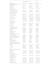
ResultsOne hundred fifty-four individuals with COPD were eligible; 89 refused to participate, and 65 were included. Four individuals withdrew during the assessments, leaving a total of 61 individuals who completed a six-month follow-up for falls (Supplementary material online 2). The anthropometric, lung function, comorbidity, medication, dyspnoea, health status, clinical postural balance, and peripheral muscle strength data are presented in Table 1. No differences were observed between FG and NFG regarding the anthropometric data, lung function, comorbidities, dyspnoea, health status, medication use, comorbidities, or muscle strength.
Baseline characteristics of the participants.
Data are presented as mean (Standard deviation) or number of individuals [ %]. Abbreviation: FG, falls group; NFG, non-falls group; BMI, body mass index; FVC, forced vital capacity; FEV1, forced expiratory volume in 1st second; TLC, total lung capacity; RV, residual volume; DLCO, diffusing capacity of monoxide of carbon; CCI, Charlson comorbidity index; mMRC, modified Medical Research Council; CAT, COPD assessment test; HADS, hospital anxiety and depression scale; HADS-A, hospital anxiety scale; HADS D, hospital depression scale; Mini-BESTest, Mini balance evaluation systems test; kgf, kilogram-force unit. % of pred., percentage of the predicted. No between-group was observed using a t-test or a Mann-Whitney test according to data normality for numerical data or a chi-square test for categorical data.
Twenty-six (42.6 %) individuals reported one or more falls, and women represented 57.7 % of FG. A total of 100 falls were recorded during the six-month follow-up period, and most falls occurred inside the house (65 %). The activities reported during the fall event, along with the main cause, are shown in the Supplementary material online 2.
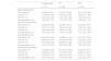
No difference between FG and NFG was detected when postural balance was assessed via the Mini-BESTest (Table 1) or via a force platform in the natural stance at rest (Table 2). However, FG presented a greater displacement in the path length and COP-AP and a higher average speed than the NFG in the natural stance post-effort condition. When postural balance was assessed at the semi-tandem position, the FG also presented greater COP-ML displacement than did the NFG. In the semi-tandem position post-effort, the FG presented greater COP displacement than did the NFG regarding all the postural balance outcomes, such as path length, COP-ML, and COP-AP (Table 2).
Postural balance assessment in individuals with COPD in different challenge positions.
Data are presented as median [interquartile range]. Abbreviation: FG, falls group; NFG, non-falls group; COP-ML, centre of pressure mediolateral displacement; COP-AP, centre of pressure anteroposterior displacement. *p < 0.05 compared with the NFG using t-test or Mann-Whitney according to data normality.
ROC curves were constructed to discriminate FG from FNG in all different postural balance conditions. The ROC curves for path length, COP-ML are presented in Figs. 1 and 2, respectively, demonstrating higher area under the curve (AUC) values in natural stance post-effort, semi-tandem, and semi-tandem post-effort. In the Supplementary material online 2, we presented the AUC for COP-AP and average speed.
 Natural stance: at rest; B) Natural stance: post-effort; C) semi-tandem: at rest; and D) semi-tandem: post-effort.")
 Natural stance: at rest; B) Natural stance: post-effort; C) semi-tandem: at rest; and D) semi-tandem: post-effort. Abbreviations: COP-ML: centre of pressure mediolateral displacement.")
Our findings demonstrated that individuals with COPD who experienced falls can be discriminated from those who did not experience falls when postural balance is assessed in more challenging conditions. No between-group differences were observed in postural balance at rest. However, FG presented greater postural balance oscillation, as assessed by the total centre of pressure (COP) and COP-AP displacements, and an increase in the average speed at the natural stance post-effort and a greater COP-ML under semi-tandem and semi-tandem post-effort conditions. Although FG and NFG presented similar lung functions, an association between lung function and postural balance was observed only under more challenging conditions.
Lung function and postural balanceMost studies involving postural balance and lung function in individuals with COPD have focused on comparing postural balance across different disease severities.37,38,39 Our results show that FG and NFG presented similar airway obstructions (FEV1), which seems similar to those reported by Castro et al.37 However, our study adds new information by demonstrating no difference in lung diffusion (gas exchange), suggesting that oxygenation does not seem relevant in distinguishing between FG and NFG. This finding could be explained by our patients presenting greater disease severity. Another possible explanation is that postural balance may be related to extrapulmonary effects, such as comorbidities and impaired muscle function. However, our findings are supported by a previous study showing that FEV1 and DLCO cannot distinguish FG and NFG.11
Postural balance conditionsThe Mini-BESTest has been proposed as a predictor of falls in individuals with COPD using the cut-off score of 22.5.32 However, in our study, both groups scored below this threshold, suggesting that, based on the Mini-BESTest, all of them have a higher probability of falls. These results suggest that the Mini-BESTest may not distinguish fall risk in individuals with more severe COPD. Our study also assessed postural balance in challenging conditions at rest and after exertion, chosen because individuals with COPD commonly present with peripheral muscle weakness and increased dyspnoea after exertion. Most previous studies evaluated postural balance in individuals with COPD at rest using force platforms. To our knowledge, none have explicitly distinguished between those who experience falls and those who do not.37,39,40
The observed differences in postural balance may be related to the varying strategies employed for maintaining posture. Balance control can be categorized into three general types: static control (maintenance of posture), proactive balance control (anticipatory adjustment), and reactive balance control (response to external perturbations).41 Importantly, individuals who experienced falls consistently attempt to compensate for postural instability via ankle and hip strategies.42 The ankle strategy is primarily responsible for maintaining postural balance while standing quietly, whereas the hip strategy compensates for larger adjustments reflected by the COP-ML differences. In our results, the ROC curve demonstrated significantly greater COP-ML displacement in semi-tandem post-effort conditions, emphasizing that the different conditions used in postural balance assessment can effectively discriminate between FG and NFG.
Our results are clinically relevant because the prevalence of falls in this study was 42.3 %, with tripping or slipping while walking being the leading cause reported. As previously reported,43 approximately, 60 % of adult falls result from unexpected perturbations during walking, such as tripping or slipping. These findings highlight the importance of our findings and reinforce the inclusion of balance training programs in challenge scenarios. To our knowledge, no such training has been specifically designed for individuals with COPD who are prone to falls.
Study limitationsThe present study has several limitations. First, we recruited only clinically stable individuals with COPD and excluded those receiving oxygen therapy. Oxygen-dependent patients may present with reduced mobility and postural balance limitations, which could bias our comparisons. Therefore, our results are not generalizable to individuals receiving oxygen therapy. Second, the force platform was used to assess postural balance. While this tool is highly accurate and is considered the gold standard for balance assessment, it is expensive and requires technical expertise, which may limit its availability in other research settings and clinical practice. This limitation suggests that more accessible methods for assessing postural balance in individuals with COPD should be explored in future studies. Third, the six-month follow-up was determined based on previous studies in individuals with COPD; however, the guidelines of falls34 in non-COPD older people recommend a period of at least 12 months. Fourth, cognitive impairment was not assessed in this study. Other factors such as age, sex, and medication consumption (antidepressant or opioid) can contribute to the risk of falls; however, no between-group difference in medication was observed. Opioid prescription was observed only in the falls group; however, only 11 % of the individuals, which does not seem relevant. Finally, the simple size calculation for the ROC curve analysis was constructed based on an expected AUC of 0.80; in contrast, our results showed an AUC of 0.65. These results may affect the accuracy of the estimated discriminative ability; however, the sample size calculation was based on detecting a between-group difference and proved adequate for the primary outcome of the study.
ConclusionOur results suggest that postural balance assessed under challenging conditions can discriminate individuals with severe to very severe COPD who have experienced a fall. Moreover, impaired postural balance contributes to an increased risk of falls, as reflected by the high prevalence of falls in this population. For clinicians, these findings underscore the importance of assessing postural balance in challenging conditions and prescribing postural balance training, particularly in this population.
The authors declare that they have no conflicts of interest to disclose.
This study was supported by the São Paulo Research Foundation (FAPESP, grant numbers 2018/17788–3, 2020/08827–5, and 2022/09628–1) and the Brazilian National Council for Scientific and Technological Development (CNPQ, grant number 312279/2018–3).
The authors thank all the individuals who participated in this research and made this possible.


