Deficits in ankle muscle strength and ankle stiffness may be present in those subjects who underwent surgical treatment for an Achilles tendon rupture. The presence of these long-term deficits may contribute to a lower performance during daily activities and may be linked to future injuries.
ObjectiveTo compare the ankle passive stiffness and the plantar flexor muscle performance in patients who underwent unilateral surgical treatment of Achilles tendon rupture with nonsurgical subjects.
MethodTwenty patients who underwent unilateral surgical treatment of Achilles tendon rupture [surgical (SU) group], and twenty nonsurgical subjects [non-surgical (NS) group] participated in this study. The ankle passive stiffness was evaluated using a clinical test. The concentric and eccentric plantar flexors performance (i.e. peak torque and work) was evaluated using an isokinetic dynamometer at 30°/s.
ResultsThe surgical ankle of the surgical group presented lower stiffness compared to the non-surgical ankle (mean difference=3.790; 95%CI=1.23–6.35) and to the non-dominant ankle of the non-surgical group (mean difference=−3.860; 95%CI=−7.38 to −0.33). The surgical group had greater absolute asymmetry of ankle stiffness (mean difference=−2.630; 95%CI=−4.61 to −0.65) and greater absolute asymmetry of concentric (mean difference=−8.3%; 95%CI=−13.79 to −2.81) and eccentric (mean difference=−6.9%; 95%CI=−12.1 to −1.7) plantar flexor work compared to non-surgical group. There was no other difference in stiffness and plantar flexor performance.
ConclusionPatients who underwent surgical repair of the Achilles tendon presented with long-term (1 year or more) deficits of ankle stiffness and asymmetries of ankle stiffness and plantar flexor work in the affected ankle compared to the uninjured side in the surgical group and both sides on the nonsurgical group.
The integrity of the Achilles tendon is important for many day-to-day activities, such as during gait, in which it has the key role of transmitting the mechanical energy generated by the contraction of the triceps surae muscles during forward body propulsion.1 Rupture of the Achilles tendon usually occurs during sports activity2 and may result in alterations of the structural and mechanical properties of the tendon.3 Surgical repair has been one of the treatment options for Achilles tendon rupture.4,5 Although the surgical treatment might restore the tendon function, some evidence indicates the presence of long-term deficits.6–8 Therefore, more studies are needed to enhance the understanding of the long-term recovery following Achilles tendon repair.
Passive ankle stiffness and the plantar flexor muscle capacity to generate strength are tissue properties that could remain affected for an extended period of time in individuals who underwent surgery due to rupture of the Achilles tendon.8 The passive stiffness reflects the ability of the structures around the joint (i.e. capsule, ligaments, fascia, and muscles) to resist passive displacement of the joint.9 The capacity of a muscle group to generate strength is commonly characterized by the variables peak torque and maximum work.10,11 Both properties, passive ankle stiffness and the capacity of the plantar flexors to generate strength, could impact the stability and movement of the ankle joint.12 Accordingly, the evaluation of these parameters in individuals having a surgical repair of the Achilles tendon would aid in the understanding of its healing process.
After the rehabilitation process and return of the individuals to their activities, it is hoped that they would exhibit tissue properties, such as stiffness and capacity to generate force, similar to asymptomatic individuals. As such, a theoretical hypothesis would be that in the long run, the surgically repaired individuals would not present deficits, or asymmetries of passive ankle stiffness and plantar flexor muscle performance compared to asymptomatic individuals with no history of tendon rupture. However, the literature suggests the possibility of deficits even after the individuals return to their activities.3 Therefore, the objectives of this study were to evaluate the passive stiffness of the ankle and the muscular performance of the plantar flexors in individuals submitted to unilateral repair of the Achilles tendon, and compare the results to individuals with no history of injury. The findings of this study may contribute to the understanding of the long-term adaptations of the tendon due to the surgical procedure.
MethodsSampleA convenience sample of forty individuals were selected and divided into two groups: surgical (SU) group and non-surgical (NS) group. The estimated sample size was calculated considering a statistic power of 80%, a probability of error type I of 0.05, and a moderate effect size (0.4).13 The SU group consisted of 20 participants with a history of unilateral rupture of the Achilles tendon, at least 12 months previously, with suture of the tendon using the Kessler and Bunnell technique (i.e. without any tendon transfer), and a history of completion of a rehabilitation program. The NS group consisted of 20 asymptomatic individuals without a history of rupture in the Achilles tendon. Exclusion criteria for both groups included current presence of musculoskeletal injuries in the lower limbs and any discomfort that could limit the performance of the tests. None of the participants were excluded. The sample characteristics are shown in Table 1. This study was approved by the Research Ethics Committee of the Mater Dei Hospital, Belo Horizonte, Minas Gerais (Number CAAE 0012.0.170.000-09). All participants signed the informed consent prior to participation.
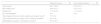
Characteristics of the subjects of surgical and nonsurgical groups.
| Surgical group (n=20) | Non-surgical group (n=20) | |
|---|---|---|
| Age (years)a | 48.7 (10.8) | 47.6 (13.1) |
| Body Mass (kg)a | 88.6 (15.4) | 79.5 (15.1) |
| Height (m)a | 1.73 (0.07) | 1.71 (0.12) |
| Sex (female/male)b | 3/17 | 8/12 |
| Time elapsed between tendon rupture and surgery (days)a | 5.5 (3.8) | – |
| Time elapsed between surgery and evaluation (months)a | 40 (10.6) | – |
| Rehabilitation (number of treatment sessions)a | 36.5 (16.1) | – |
Initially, anthropometric measures, such as body mass and height using a digital scale with altimeter (Filizola® SA, São Paulo, Brazil), were obtained. Following, clinical testing of passive ankle stiffness14 and evaluations of the plantar flexors performance with the isokinetic dynamometer (Biodex System 3 Pro Biodex Medical Systems Inc., Shirley, USA)15 were then conducted. Testing was done to both limbs of all subjects.
Passive stiffness of the ankleThe passive ankle stiffness was evaluated through a clinical test.14 This test had been validated with direct measurement of passive stiffness using the isokinetic dynamometer in a previous study.14 Therefore, it could be used as a clinical test to index the passive stiffness of the ankle.14 To perform this clinical test, the protocol recommended by Araújo et al.14 was used. Each participant was positioned in prone with the knee flexed at 90° for each leg to be tested.14 In this position, markers were placed on the fibular head and lateral malleolus, and then a line was drawn using a ruler joining these two points, which afterwards served to align the fixed arm of the goniometer (Fig. 1A).14 Then, the evaluator conducted a viscoelastic accommodation of the periarticular tissues of the ankle obtained through a series of five sequential movements of dorsiflexion to its maximum amplitude.14 Next, an ankle weight of 2kg was placed 8cm from the lateral malleolus to generate tension in the plantar flexors (Fig. 1A).14 The use of an ankle weight was a procedure recommended by the authors who had validated the test, since the torque produced only by the mass of the foot was insufficient to tension the plantar flexors (i.e. the position of the foot could remain unchanged even with the action the gravity favoring dorsiflexion).14 The position taken by the ankle joint immediately after placing the ankle weight was measured using the goniometer (CARCI®, São Paulo, Brazil). The stationary arm of the goniometer was aligned with a line joining the lateral malleolus to the fibular head, and the moveable arm was parallel to the surface of the foot (Fig. 1A).14 Three measurements were performed on each ankle joint, starting with the participant's preferred lower limb. Each participant was asked to avoid contracting the muscles in the lower limb being investigated so that there was minimal or no influence on the measurement.14 The evaluator palpated the tibialis anterior, triceps surae, and hamstrings muscles during the test to verify the lack of muscular contraction.14 If the participant was contracting any of these muscles, the test was discarded and repeated. This test was performed by a single examiner who showed excellent test–retest reliability16 (intraclass correlation coefficient – ICC3,2=0.84 (95% CI=−0.14 to 0.98) standard error of measurement – SEM=2.73), evaluated in a pilot study with 10 volunteers conducted one week apart.

(A) Clinical test of ankle passive stiffness: Participant was positioned in prone with the knee flexed at 90°. An ankle weight of 2kg was placed 8cm from the lateral malleolus. The position taken by the ankle was measured using the goniometer (the moveable arm was aligned parallel to the surface of the foot). (B) Evaluation of ankle plantar flexor performance at the isokinetic dynamometer.
Initially, all volunteers warmed up by walking at a self-selected fast speed on the ground for five minutes. Next, they seated in the chair of the isokinetic dynamometer, with belts over the trunk, pelvis, and thigh for stabilization. The seat back tilt was set at 70° and each participant's distal thigh was supported over the device's limb-support pad so that the knee remained flexed between 30° and 40°. This range was assured by the evaluator using the goniometer. The dynamometer's axis of rotation was aligned with the lateral malleolus and the barefoot was strapped to the footplate of the ankle attachment of the isokinetic dynamomater so that the plantar surface of the foot was fully supported on this base of support that was attached to the dynamometer (Fig. 1B).
The protocol17,18 consisted of concentric and eccentric evaluations of the plantar flexors muscles, within a range of 10° of dorsiflexion and 20° of plantar flexion, repeated five times at a 30°/s velocity. Initially, the participants were familiarized with the system by performing five repetitions using submaximal contractions.19 The assessment was initiated with the non-surgical lower limb of the participants in the SU group, and with the dominant lower limb (i.e. the limb preferred when kicking a ball) of the participants in the NS group. This order was established to ensure security to the volunteers during the test, especially for the SU group. During the test, participants were instructed to perform with maximal force. Standardized verbal encouragement was provided by the researcher to ensure maximum force generated by the subjects.20
The reliability and validity of the isokinetic dynamometer had been demonstrated in the literature.15 In addition, the test using the isokinetic dynamometer was conducted by only one evaluator with extensive experience with the use of the equipment. The test–retest reliability of the protocol was determined in a pilot study using 10 volunteers, evaluated at an interval of one week apart that showed excellent16 reliability results for the outcome variables studied (ICC3,2=0.92 to 0.94; (95% CI=0.74–0.98) SEM=5.85–12.05).
Data reductionInitially, to enable data analysis, the output from the lower limbs of the participants from both groups were paired such that: the non-dominant lower limb data of the NS group were considered as corresponding to the surgical lower limb data of the SU group; and the dominant lower limb data of the NS group corresponding to non-surgical lower limb data of the SU group.
The variable passive stiffness of the ankle was determined in degrees from the average of the three values obtained for each lower limb through the clinical passive stiffness test. In addition, the absolute asymmetry of this variable was calculated from the difference between the lower limb side with higher value of passive stiffness and the lower limb side with lower stiffness value.
The performance of the plantar flexor muscles was analyzed using the concentric and eccentric peak torque normalized by body weight, as well as the concentric and eccentric maximum work performed in one repetition, also normalized by body weight. In addition, the absolute asymmetries of the peak torque and maximum work were analyzed. The asymmetry values were obtained from the report of the isokinetic dynamometer software, but without considering the direction (absolute asymmetry).21,22 The asymmetry calculation was performed according to the equation: Asymmetry=((Non-surgical or dominant side−Surgical or non-dominant side)/Non-surgical or dominant side)×100.
Statistical analysisThe assumptions of normality and homogeneity of variance were checked and confirmed prior to the inferential tests. Mixed analysis of variance (ANOVA) with one independent factor (SU and NS groups) and with one repeated measure factor (non-surgical/dominant lower limb and surgical/non-dominant lower limb) was used to analyze the effects of group, lower limb and the interaction effect of lower limb×group on the passive stiffness of the ankle. Independent t-test was performed to verify if the absolute asymmetry of passive stiffness was different between groups.
Multivariate analysis of variance (MANOVA) with a mixed model was used to evaluate the effect of group, lower limb, and the interaction effect of group×lower limb on the muscle performance variables (i.e. concentric peak torque, eccentric peak torque, maximum concentric work and maximum eccentric work of the plantar flexors of the ankle normalized by body weight). In addition, MANOVA was also conducted to verify the effect of group on the variables of absolute asymmetry of muscle performance. One-way ANOVA were used to identify possible differences observed by both MANOVA. For all the analyses presented, a type I error probability of 0.05 was considered. All data were analyzed using the software IBM SPSS version 20.0 (IBM Corp.).
ResultsPassive ankle stiffnessThe ANOVA revealed no main group effect in the passive ankle stiffness (F1,38=1.760, p=0.19; η2=0.044; mean difference=2.190; 95% CI −1.16 to 5.54). However, a main lower limb effect (F1,38=8.129, p=0.01, η2=0.176; mean difference=2.130; 95% CI=0.62–3.64) was observed in which the passive stiffness was reduced (i.e. greater angular displacement) in the surgical/non-dominant lower limb (9.0±0.9°) compared to the non-surgical/dominant lower limb (6.8±1.0°). In addition, there was an interaction effect lower limb×group (F1,38=4.937, p=0.03; η2=0.115). Four contrasts were conducted to investigate this interaction effect. The surgical lower limb of the SU group presented reduced stiffness compared to non-surgical lower limb (t19=3.096, p=0.01; mean difference=3.79, 95% CI=1.23–6.35). The surgical lower limb of the SU group presented also reduced stiffness compared to non-dominant lower limb of the NS group (t38=−2.212; p=0.03; mean difference=−3.86; 95% CI=−7.38 to −0.33). The non-surgical lower limb of the SU group was not different from the dominant lower limb of the NS group (t38=−0.284; p=0.78; mean difference=−0.54; 95% CI=4.35–3.28). In addition, the dominant lower limb of the NS group was not different from the non-dominant lower limb of the NS group (t19=0.548, p=0.59; mean difference=0.47; 95% CI=1.32–.26).
The independent t-test showed a difference between groups in the absolute stiffness asymmetry of the ankle (t38=−2.689; p=0.01; mean difference=−2.630; 95% CI=−4.61 to −0.65). The SU group (5.6±3.6°) was more asymmetric than the NS group (2.9±2.4°).
Performance of the ankle plantar flexorsThe MANOVA showed no main group effect (F4,35=1.943, p=0.13, η2=0.182; range of mean differences=2.088–28.345), lower limb effect (F4,35=1.989, p=0.12; η2=0.185; range of mean differences=2.372–10.615), or interaction effect of lower limb×group (F4,35=0.317, p=0.87, η2=0.035) on the muscular performance variables of the plantar flexors. The mean and standard deviation of the muscular performance variables of the plantar flexors, according to the group and lower limb, are shown in Table 2.
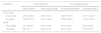
Peak torque (Nm/kg) and work (J/kg) of ankle plantar flexor muscles of each limb and for surgical and nonsurgical groups.
| Variables | Surgical group | Non-surgical group | ||
|---|---|---|---|---|
| Surgical limb | Non-surgical limb | Non-dominant limb | Dominant limb | |
| Peak torque | ||||
| Concentric | 179.4 (48.4) | 188.9 (46.1) | 202.9 (45.7) | 214.7 (48.2) |
| Eccentric | 202.0 (53.8) | 211.8 (48.4) | 229.4 (46.0) | 241.0 (48.8) |
| Work | ||||
| Concentric | 45.1 (16.6) | 47.8 (12.0) | 48.2 (14.0) | 50.3 (14.8) |
| Eccentric | 58 (19.1) | 60.9 (13.9) | 60.2 (15.9) | 62.8 (16.5) |
Data presented as mean (standard deviation).
In relation to the asymmetry variables of muscle performance, the MANOVA identified differences between the SU and NS groups (F4,35=4.258, p=0.01, η2=0.327). The SU group was more asymmetric than the NS group in the absolute asymmetry variables of concentric work (F1,38=7.010, p=0.01, η2=0.156) and eccentric work (F1,38=7.022, p=0.01, η2=0.156). There was no difference between groups for the absolute asymmetry variables of concentric torque (F1,38=3.478, p=0.07, η2=0.084) and eccentric torque (F1,38=2.003, p=0.17, η2=0.050). The mean, standard deviation, mean difference, and confidence intervals of absolute asymmetry variables are shown in Table 3.
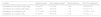
Absolute asymmetry (%) of ankle plantar flexor muscle performance of surgical and nonsurgical groups.
| Variable | Surgical groupa | Non-surgical groupa | Mean difference | 95% CI of difference |
|---|---|---|---|---|
| Asymmetry of concentric peak torque | 12.9 (9.7) | 8.1 (5.8) | −4.8 | −9.92 to 0.32 |
| Asymmetry of eccentric peak torque | 12.0 (9.3) | 8.6 (5.6) | −3.4 | −8.31 to 1.51 |
| Asymmetry of concentric work | 17.3 (11.8) | 9.0 (8.0) | −8.3 | −13.79 to −2.81 |
| Asymmetry of eccentric work | 15.9 (9.1) | 9.0 (7.0) | −6.9 | −12.1 to −1.7 |
The present study observed that, even with at least one year post surgery, individuals who had an Achilles tendon repair, had changes in the passive stiffness of ankle and performance of the plantar flexor muscles on the surgical leg. Specifically, reduced passive stiffness in the surgical ankle, greater stiffness asymmetry, and greater asymmetry on the capacity to generate concentric and eccentric work of the plantar flexors was observed in individuals who had reconstruction of the Achilles tendon, compared to individuals with no history of a similar surgical procedure.
The results of this study indicated that the surgical side of the SU group had reduced ankle passive stiffness (10.8±4.7°) compared to the non-surgical side of the same group (7.1±5.4°) or with the corresponding side of the NS group (7.0±6.2°). Moreover, it was observed that the SU group had increased absolute asymmetry of ankle passive stiffness (5.6±3.6°) compared to the NS group (2.9±2.4°). These results, contrary to the hypothesis of the study, indicated that even after one and a half years of surgery, the passive stiffness was different from individuals with no history of injury. After rehabilitation and return to the usual activities, it would be expected that the tension stimuli arriving at the periarticular tissue would favor the injured tissue acquiring similar properties observed in a non-surgical ankle. In support to this argument, Don et al.23 observed that after two years of surgery for Achilles tendon rupture, the ankle passive stiffness values were similar between the surgical and non-surgical limbs. However, there is evidence in the literature of reduced stiffness on the surgical side even after two years of surgery.3 Since the joint stiffness is associated with muscular trophism,24 a possible explanation for the reduced stiffness on the surgical limb, as observed in this study, would be the small cross-sectional area of the plantar flexors on the limb submitted to surgery. However, as the cross-sectional area was not assessed in this study, this explanation remains speculative.
The reduced stiffness on the surgical side indicated the decreased capacity of periarticular tissues to resist displacement of the joint when an external force was applied. This finding was of particular interest because a joint with reduced levels of stiffness might be more vulnerable to the effects of an external disturbance, since there is a close relationship between stiffness and joint stability.12,24,25 Moreover, the reduction in ankle stiffness levels could favor excessive movement at this joint and increase the demand for muscle activation during functional activities (e.g. gait)26,27 and in other structures of the musculoskeletal system. Accordingly, the permanent stiffness deficits (reduced stiffness on the surgical limb) could lead to a less stable joint and consequently, could be more prone to new injuries. Future studies are needed to investigate this proposition.
Since the passive stiffness of a joint could be influenced by the muscular trophism,24 it was possible that reduced levels of ankle passive stiffness, observed on the surgical limb, might have been followed by decreased force-generating capacity of the ankle muscles; in particular, the triceps surae muscles. However, confirming the hypothesis of the study, no main effects of group and lower limb or interaction effect for the variables of muscle performance were observed. The literature also showed contradictory results in relation to the different variables that characterize the triceps surae muscle performance in individuals having a surgical reconstruction.23,28 Possible reasons for the different results reported are the surgical procedures performed or the evaluation methods used in the studies.
The individuals from the SU group had greater asymmetry between lower limbs in the concentric and eccentric work of plantar flexors when compared to the individuals of the NS group. In other words, the plantar flexors of the participants of the SU group showed differences between the limbs (absolute and not relative difference) in the ability to generate torque throughout the range of motion. It has been argued in the literature that asymmetries in the muscular performance variables could arise due to the differences in patterns used by the lower limbs during functional activities23 due to incomplete rehabilitation programs,29 or due to specific functional demands.29 Although the literature supported that these factors could be the cause for the development of asymmetries, the possible reasons for the asymmetries observed in the present study should be viewed with caution, since these factors were not investigated in this study.
The results obtained in this study indicated that passive stiffness levels of the ankle and work of the plantar flexors were different between lower limbs of individuals having an Achilles tendon repair; even after a long postoperative period. This study did not investigate how the SU group individuals performed their rehabilitation program. However, the lack of treatment information might be considered a limitation of this study since knowledge of the rehabilitation process would help to better understand the results. Another limitation of this study was the lack of the functional status of the participants, which could provide information about their return to usual activities and thus, also contribute to the interpretation of the findings. In addition, the absence of a matching procedure between groups is a limitation of this study. Despite that, the groups had similar mean values of age, body mass and height (Table 1). The results of this study provide evidence of the permanence of deficits and asymmetries in tissue properties, such as passive stiffness and muscle work, even after a long period, in individuals submitted to Achilles tendon repair.
ConclusionsThe results of this study conducted in individuals submitted to unilateral Achilles tendon repair at long term, showed reduced passive stiffness in the surgical ankle, in addition to absolute asymmetry of the passive stiffness of the ankle and the capacity to generate concentric and eccentric work of the plantar flexors.
Conflicts of interestThe authors declare no conflicts of interest.
The authors thank Juliana Alves de Andrade, João Murilo Magalhães, Estela Vieira and Breno Lopes for helping in data collection and the Brazilian government agencies: CNPq, FAPEMIG and CAPES.