


This study had three aims: (1) to evaluate the relationships between the paretic knee extensor muscle strength and global lower-limb strength in individuals who had suffered a sub-acute/chronic stroke, (2) to determine whether global lower-limb strength, sex, body mass index, or age could predict knee extensor muscle strength, and 3) to investigate whether the results obtained via a Modified Sphygmomanometer Test (MST) would be similar to those obtained using a hand-held dynamometer.
MethodsThis was a cross-sectional study, performed at a research laboratory, at participants’ homes, or at outpatient clinics. Forty-two individuals with a sub-acute stroke and 45 individuals with a chronic stroke participated. Maximum isometric strength of the paretic lower-limb muscles (i.e. hip, knee, and ankle flexors/extensors, hip abductors) was measured using the MST and a hand-held dynamometer.
ResultsSignificant and high correlation coefficients were found between knee extensor muscle strength and global lower-limb strength as measured by the combined strength values of 6 lower limb muscle groups in individuals with sub-acute (0.81≤r≤0.88; p<0.05) and chronic (0.82≤r≤0.85; p<0.05) stroke. Step-wise multiple regression analysis revealed that only global lower-limb strength was retained in the model and accounted for 66–78% and 67–72% (p<0.001) of the variance in knee extensor muscle strength at the sub-acute and chronic phases post-stroke, respectively. The results obtained via the MST were similar to those obtained using the hand-held dynamometer.
ConclusionParetic knee extensor muscles strength, assessed using a MST or a hand-held dynamometer, indicates global lower-limb strength in individuals with a sub-acute or chronic stroke.
Stroke is the second leading cause of number of years living with disability worldwide.1 One common post-stroke impairment is lower-limb muscle weakness, which is associated with functional limitations.2–7 In addition, strength deficits of the paretic lower limb were the physical impairments that best explained the restricted participation in life situations of individuals who had suffered a stroke.8 Therefore, the assessment of muscle strength of individuals with stroke is considered relevant and recommended3 and is commonly used in both clinical and research settings.
It has been shown that the strength measurement of a single muscle group is a fast, simple, and objective way to describe the strength of the entire limb.9–11 According to the results of a systematic review, the knee extensors were the lower-limb muscle group that was most frequently measured using a dynamometer in stroke individuals.12 Knee extensor muscle strength has been shown to reflect overall lower-limb strength of healthy individuals,13,14 and previous studies have shown that the strength of the paretic knee extensor muscles was significantly correlated with other muscle groups of the paretic lower limb following a stroke.15–17 However, the current available data are incomplete since these relationships were only investigated for the hip flexors, knee flexors, and ankle dorsiflexors.15–17 In addition, the results of these studies cannot be generalized to the stroke population since only individuals in the acute phase were evaluated. Furthermore, all previous studies employed a hand-held dynamometer to obtain strength measurements.15–17 However, despite the hand-held dynamometer being a reliable, and valid instrument for assessing isometric muscle strength,18 its relatively high cost may decrease its use in some areas. Therefore, the Modified Sphygmomanometer Test (MST) is an alternative instrument that has been used to obtain objective muscle strength measurements in clinical settings.19,20 The MST showed adequate reliability and validity (when compared against the hand-held dynamometer) for muscle strength measurements of individuals in the sub-acute and chronic phases post stroke.19,20 In addition, to use the MST, only a sphygmomanometer is required, which is both inexpensive and commonly used by most healthcare professionals.10,19,20 Nevertheless, the relationships between MST measures of the knee extensor muscles and global lower-limb strength in individuals with stroke have not been investigated.
Therefore, the objectives of this study were threefold: (1) to evaluate the relationships between the paretic knee extensor muscle strength and global lower-limb strength as measured by the combined strength values of 6 lower limb muscle groups in individuals with sub-acute/chronic stroke; (2) to determine whether global lower-limb strength, sex, body mass index, or age could predict knee extensor muscle strength; and (3) to investigate if the strength measures obtained via MST were similar to those measured using the hand-held dynamometer.
MethodsParticipantsIndividuals who have suffered a stroke were recruited from the community. These individuals were included in the study if they had a clinical diagnosis of stroke (sub-acute: 3–6 months; chronic: >6 months post-stroke) and were ≥20 years of age. Those who were excluded were individuals with cognitive impairment (as determined by education-specific cut-off values on the Mini-Mental State Examination21 or were unable to understand verbal commands)22; were unable to perform the tests; and/or had other chronic diseases or pain during the tests. All participants provided written consent for participation in this cross-sectional study, which received approval from the institutional ethical review board (Universidade Federal de Minas Gerais-UFMG, Belo Horizonte, Minas Gerais, Brazil, #0492.0.203.000-10).
The lower-limb section of the Fugl-Meyer Motor Assessment Scale was used to assess motor function of the lower limb. This scale has presented adequate validity and reliability in stroke individuals.23 Motor impairments were classified as mild (>29 points), moderate (23–29 points), moderately severe (18–22 points), and severe (<17 points).24
Outcome measuresThe isometric strength of the paretic knee extensors and the global strength as measured by the combined strength values of 6 lower limb muscle groups (i.e. hip and ankle flexors/extensors, hip abductors, and knee flexors) were measured using a hand-held digital Microfet2® dynamometer (Hoggan Health Industries, Salt Lake City UT, USA) and the MST (DuraShock™ Tycos® aneroid sphygmomanometer model DS-44; Welch Allyn Inc, Skaneateles Falls, NY, USA). The sphygmomanometer was adapted using the bag method.19,20,25 To adapt the sphygmomanometer using the bag method the inflatable part of the entire external Velcro band of the sphygmomanometer was removed, folded into three equal sections, placed in an inelastic zippered cotton bag, and calibrated according to recommended procedures.19,20,25
ProceduresPrior to the testing, the examiner underwent a training period consisting of performing one-week of muscle strength measurements to the muscle groups to be tested under the supervision of other physical therapists who were not involved in the testing. The tests were conducted on a single day by a trained physical therapist. Each participant had all muscles tested in a single day. The reading and recording of the strength measurements were performed by another examiner. No communication was allowed between the examiners regarding the obtained data. Clinical and demographic data were collected, followed by the muscle strength measurements (i.e. dynamometry and MST). Before the measurements were taken, the order of the instruments (hand-held dynamometer or modified sphygmomanometer) was randomized. A 5-min rest interval was allowed between the measurements with each device.10
The data on the strength of the paretic limb were obtained in the following order: global lower-limb strength (hip and ankle flexors/extensors, hip abductors, and knee flexors) and knee extensor strength. However, not all muscle groups of some participants could be assessed, because they were unable to generate sufficient isometric force.
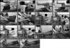
Positioning, manual stabilization, place of resistance applications, and verbal encouragement were standardized and followed previously described protocols.19,20 The muscle strength measurement procedures were the same for the hand-held dynamometer and the MST. For assessment of the hip and ankle flexors/extensors and hip abductors, the individuals were tested in the supine position. For measurements of the knee flexors/extensors, the participants were tested in the seated position.19,20 Participant and device positioning was as follows (zero degrees meant that the joint was in neutral position, i.e., neither in flexion nor in extension) (Fig. 1):
- -
Hip flexors/extensors: The hip and knee joints were flexed to 90° and the test apparatus was positioned proximal to the anterior/posterior surface of the knee.
- -
Hip abductors: The hip and knee joints were maintained at 0° and the apparatus was positioned on the lateral surface of the thigh proximal to the knee.
- -
Ankle dorsi/plantar flexors: The hip and knee joints were at 0° and the apparatus was positioned on the dorsal/plantar surfaces of the foot proximal to the metatarsophalangeal joints.
- -
Knee flexors/extensors: The back and feet were unsupported, the hip and knee joints were flexed to 90°, and the apparatus was positioned on the posterior/anterior surface of the leg proximal to the ankle.

Measurement of muscle strength using a hand-held dynamometer (A – hip flexors, C – hip extensors, E – hip abductors, G – knee flexors, I – knee extensors, K – ankle dorsiflexors, M – ankle plantar flexors) and a modified sphygmomanometer test (B – hip flexors, D – hip extensors, F – hip abductors, H – knee flexors, J – knee extensors, L – ankle dorsiflexors, N – ankle plantar flexors).
Demonstration and familiarization procedures were performed with both instruments before measurements were taken. For each contraction, each participant was instructed to perform a maximum isometric contraction for 5s and the peak force was recorded. After familiarization, only one trial was performed, based upon findings from previous studies.20,21,26,27 If the examiner observed any compensatory movement by the participants, such as using the upper limb for stabilization purposes or moving the trunk posteriorly during the knee extensor measurement, another measurement was taken.20,21,26,27 However, this was not frequent, and no data was lost because of that. The force exerted on the modified sphygmomanometer was determined by the pressure gauge reading in 2 – mmHg increments. Before each MST measurement, the examiner ensured that the sphygmomanometer was pre-inflated to 20mmHg.10
Statistical analysisThe sample size of 14 individuals per group was determined to be the minimum sample size needed for the study using MedCalc® software (version 12.7.5 for Windows; Ostend, Belgium) considering a power of 0.80, r=0.69 (moderate correlation),28 and α=0.05. To correctly understand the relationships between two variables, that is, for correlation analyses, it is desirable to include as wide a range of muscle strength values as possible.29 Therefore, to achieve wide variability of muscle strength measures, the recruitment considered two age groups (20–59 years of age and ≥60 years of age) and both sexes (i.e. males and females). Thus, a sample of at least 28 individuals at the sub-acute and chronic phases was required as a minimum sample.
Descriptive statistics and normality tests (using the Shapiro–Wilk Test) were performed for all variables. Global strength of the paretic lower limb was determined by the sum of the strength values of the hip and ankle flexors/extensors, hip abductors, and knee flexors taken using a hand-held dynamometer and a MST. Pearson correlation coefficients were calculated to determine the magnitudes and directions of the correlations between the strength values (from the MST and dynamometer) of the knee extensor muscles and global strength of the paretic lower limb. When significant, the correlations were classified as follows28: very low: ≤0.25; low: 0.26–0.49; moderate: 0.50–0.69; high: 0.70–0.89; and very high, 0.90–1.00. Four step-wise multiple regression models (two for the sub-acute and two for the chronic phases) were employed to investigate whether sex, age, body mass index, or global lower-limb strength would predict knee extensor muscle strength measured using the MST and the dynamometer. It is worth noting that, for the model where the dependent variable was knee extensor muscle strength evaluated by using the hand-held dynamometer, the global strength of the paretic lower limb, also measured by using the hand-held dynamometer, was considered as a predictive variable, maintaining similarities with the MST measurements. All assumptions regarding the regression analyses were verified and adequately met (α=5%).
ResultsFrom 659 potential participants, 326 could not be contacted due to inaccurate contact information. From the contacted participants, the main reasons for exclusion were refusals (n=80), inability to walk (n=40), transportation issues (n=25), lack of time (n=24), and other non-stroke related conditions (n=23). Eighty-seven individuals with stroke were evaluated: 42 in the sub-acute phase (26 men, 16 women; mean age, 62 years (SD 13); time post-stroke, 3.7 months (SD 0.9)) and 45 in the chronic phase (24 men, 21 women; mean age, 56 years (SD 13); mean time post-stroke, 92 months (SD 74)). Their clinical and demographic characteristics are described in Table 1, whereas the strength values of the paretic lower limb are reported in Table 2. Six individuals (two men and four women, who had a mean age of 60 years (SD 16) and a mean time since stroke of 3.5 months (SD 0.8)) at the sub-acute post-stroke phases could not have all muscle groups assessed, due to difficulty in generating isometric force. All of these individuals had ischemic stroke and lower-limb impairments (three severe, two moderate, and one mild impairments), according to the Fugl-Meyer scale scores. The strength of the ankle dorsiflexor/plantar flexor and knee flexor muscles was not assessed in some participants with sub-acute stroke. Eleven individuals (six men, five women, who had a mean age of 55 years (SD 10) and a mean time since stroke of 80 months (SD 74)) at the chronic post-stroke phases could not also have all muscle groups assessed, due to difficulty in generating isometric force. All of these individuals had ischemic stroke and lower-limb impairments (three severe, three moderately severe, four moderate, and one mild impairments). The strength of the following muscles groups of some participants with chronic stroke was not assessed, as follows: ankle dorsiflexors in six individuals, plantar flexors in five, knee flexors in four, and hip flexors/extensors in three individuals. A measure of strength of the knee extensor muscles of one participant in the chronic phase post-stroke was excluded, because it was above the upper capacity of measurement of the modified sphygmomanometer.
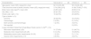
Demographic and clinical characteristics of the participants to determine whether knee extensor muscles strength indicates global lower limb strength in individuals suffering from a stroke.
| Characteristics | Sub-acute (n=42) | Chronic (n=45) |
|---|---|---|
| Age (years): mean (SD); range [min–max] | 62 (13) [35–85] | 56 (13) [30–86] |
| Time since the onset of stroke (months): mean (SD); range [min-max] | 3.74 (0.86) [3–6] | 91.98 (73.86) [7–370] |
| Body mass index (kg/m2): mean (SD) | 24.85 (4.40) | 26.95 (5.16) |
| Sex: Men, n (%) | 26 (61.9%) | 24 (53.3%) |
| Paretic side: right, n (%) | 18 (42.9%) | 22 (48.9%) |
| Type of stroke: n (%) | ||
| Ischemic | 39 (92.9%) | 34 (75.6%) |
| Hemorrhagic | 2 (4.8%) | 6 (13.3%) |
| Both ischemic and hemorrhagic | 1 (2.4%) | 3 (6.7%) |
| Not reported | – | 2 (2.2%) |
| Lower limb Motor Impairment (Fugl-Meyer Scale) (score: 0–34)24: n (%) | ||
| Mild motor impairments (>29) | 31 (73.8%) | 21 (46.7%) |
| Moderate motor impairments (23–28) | 6 (14.3%) | 16 (35.6%) |
| Moderately severe motor impairments (18–22) | 1 (2.4%) | 5 (11.1%) |
| Severe motor impairments (<17) | 4 (9.5%) | 3 (6.7%) |
SD, standard deviation.
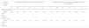
Descriptive statistics regarding the measures of knee extensor muscles and global strength of the paretic lower limb for individuals with subacute and chronic stroke.
| Strength measure | Instruments | n | Mean (SD) | 95% CI | Range [min–max] |
|---|---|---|---|---|---|
| Sub-acute phase | |||||
| Knee extensors | Modified sphygmomanometer (mmHg) | 42 | 166.14 (60.74) | 154.46–194.87 | 30–280 |
| Hand-held dynamometer (kg) | 42 | 10.09 (4.72) | 9.24–12.40 | 1.8–19.1 | |
| Global lower limb strength | Modified sphygmomanometer (mmHg) | 36 | 765.61 (209.76) | 694.64–836.58 | 272–1140 |
| Hand-held dynamometer (kg) | 36 | 48.83 (16.86) | 43.12–54.54 | 15.20–88.9 | |
| Chronic phase | |||||
| Knee extensors | Modified sphygmomanometer (mmHg) | 44 | 163.182 (52.99) | 151.50–186.92 | 60–274 |
| Hand-held dynamometer (kg) | 45 | 13.12 (6.18) | 11.18–15.33 | 2.8–27.3 | |
| Global lower limb strength | Modified sphygmomanometer (mmHg) | 34 | 754.35 (216.42) | 676.68–832.53 | 296–1206 |
| Hand-held dynamometer (kg) | 34 | 56.95 (19.62) | 50.02–64.15 | 21–105 | |
SD, standard deviation; CI, confidence interval.
Strength of the paretic knee extensor muscles showed significant and high correlations with those of global lower-limb strength obtained using both the MST (0.85≤r≤0.88) and hand-held dynamometer (0.81≤r≤0.82) for both individuals in the sub-acute and chronic phases (Table 3).
Correlation coefficients between the strength measurements (i.e. MST and hand-held dynamometer) of the knee extensor muscles and global strength of the paretic lower limb strength as measured by the combined strength values of 6 lower limb muscle groups for individuals with subacute and chronic stroke.
| Variables | n | Knee extensor strength (MST) | n | Knee extensor strength (Hand-held dynamometer) |
|---|---|---|---|---|
| Sub-acute phase | ||||
| Global lower-limb strength | 36 | r=0.88; p<0.001 | 36 | r=0.81; p<0.001 |
| Chronic phase | ||||
| Global lower-limb strength | 33 | r=0.85; p<0.001 | 34 | r=0.82; p<0.001 |
MST, modified sphygmomanometer test; r, Pearson correlation coefficient.
Table 4 shows the results of the regression analysis for both sub-acute and chronic stroke sub-groups and the data obtained using the MST and the hand-held dynamometer. The regression models for the data obtained using MST showed results similar to those obtained using the hand-held dynamometer. From the four predictors, only global strength of the paretic lower limb was retained in all models and explained 66–78% (p<0.001) of the variance of knee extensor muscle strength for the participants in the sub-acute and chronic phases.
Results of the regression models used to indicate whether knee extensor muscles strength indicates global lower-limb strength in individuals who have suffered a stroke.a
| Predictor variable model | Dependent variable knee extensor strength (MST) | Dependent variable knee extensor strength (hand-held dynamometer) | ||||||||||
|---|---|---|---|---|---|---|---|---|---|---|---|---|
| B | SE | β | R2 | F | p | B | SE | β | R2 | F | p | |
| Sub-acute phase | ||||||||||||
| Global lower-limb strength | 0.25 | 0.02 | 0.88 | 0.78 | 119.19 | <0.001 | 0.22 | 0.03 | 0.81 | 0.66 | 65.50 | <0.001 |
| Chronic phase | ||||||||||||
| Global lower-limb strength | 0.19 | 0.02 | 0.85 | 0.72 | 80.01 | <0.001 | 0.25 | 0.03 | 0.82 | 0.67 | 67.65 | <0.001 |
a All of the step-wise regression models included the following input variables: sex, age, body mass index, or global strength of the paretic lower limb. MST, modified sphygmomanometer test; B, regression coefficient; SE, standard error of regression coefficient; β, standardized regression coefficient; R2, coefficient of determination; F, F-statistic; p, significance level.
The findings of the present study showed that the paretic knee extensor muscles strength was significantly, positively, and strongly correlated with the global lower-limb strength as measured by the combined strength values of 6 lower limb muscle groups in individuals with sub-acute and chronic stroke. In addition, only global lower-limb strength was retained in the regression models and had significant predictive values. Finally, the results of the statistical analysis obtained using the MST were similar to those obtained when using the hand-held dynamometer for individuals in the sub-acute and chronic phases of stroke.
Significant and strong correlations were found between the knee extensor muscle strength and global lower-limb strength as measured by the combined strength values of 6 lower limb muscle groups in individuals in the sub-acute (0.81≤r≤0.88) and chronic (0.82≤r≤0.85) phases. These results corroborate those of a previous review of the literature that claimed that it was valid to use strength measurements from fewer muscle groups to characterize the strength of an entire limb.7 In addition, these results agree with those of a similar study of healthy individuals, which also found significant correlations between dynamometric measurements of strength of the knee extensors and those of other lower limb muscles (i.e. hip flexors, ankle dorsiflexors).13 However, the magnitude of the correlations ranged from low to high (0.41≤r≤0.73).13 Another similar study, which included only healthy elderly individuals, also found significant correlations of moderate to high magnitudes (0.51≤r≤0.97)14 between the knee extensor strength and that of other lower limb muscles (i.e. hip flexors/abductors, knee flexors, and ankle dorsiflexors). The three studies that examined the relationships between dynamometric measurements of strength of the paretic knee extensors and other muscle groups (i.e. hip and knee flexors, ankle dorsiflexors) of the paretic lower limb in individuals suffering from an acute stroke also reported significant correlations of moderate to high magnitudes (0.66≤r≤0.87).15–17
In the present study, the results of the multiple regression analyses showed that among sex, age, body mass index, or global strength of the paretic lower limb, the latter was a significant predictor of the variance in paretic knee extensor muscle strength and explained 66–78% of the variance in paretic knee extensor muscle strength for both stroke sub-groups. The results of some of the previously cited studies that investigated the relationships between measurements of strength of the knee extensors and those of other muscle groups of the lower limbs were based on univariate analyses, that is, correlation coefficients,15,16 although none employed regression analyses. Although regression analysis is considered better than factor analysis,29 some studies used factor analysis to explore these relationships.13,14 The strength of individual muscle groups explained 59–87% of the variance of global lower-limb strength.13,14
The similar statistical results obtained through the present study regarding the strength obtained using the hand-held dynamometer and the MST support previous findings that claimed adequate validity of the MST for measuring the muscle strength of individuals who have suffered a stroke.19,20 Therefore, besides demonstrating adequate values of test-retest/inter-rater reliabilities and criterion-related validity,19,20 the MST may also be used to characterize global lower-limb strength by assessing only one muscle group (i.e., knee extensors), in individuals in the sub-acute and chronic phases of stroke. Given these research results, associated with low cost, portability, and simplicity, the MST use in clinical practice is compelling.
This study does have some limitations. First, the participants were selected using a convenience sample and few had severe lower-limb motor impairments. Thus, the findings might not be representative of the entire stroke population. In addition, not all muscle groups of some participants could be assessed, because they were unable to generate sufficient isometric force. However, this probably did not affect the results since we recruited a sample bigger than the minimum required according to the sample size calculation in an order to obtain sample heterogeneity regarding strength values. This was done because one important assumption in correlation analysis is sample heterogeneity regarding the outcome of interest.29 The results indicated that a measurement of strength of only one muscle group (i.e. knee extensors) could be used to reflect global lower-limb strength. Nevertheless, if the aim is to obtain information about the strength of a specific muscle group, its assessment is certainly necessary. In addition, one limitation of the muscle strength measurement with the MST was that some individuals might be stronger than the instrument's reading capacity. However, this limitation is mostly found when the MST is employed with the bag method.27 Therefore, future studies should also investigate if the results found in the present study are also true with the use of other adaptations of the sphygmomanometer to perform the MST. Moreover, not all muscle groups of the lower limb, such as the hip internal/external rotators, were assessed and future studies should include these measurements. Future studies should also investigate whether other lower limb muscle groups could indicate global lower-limb strength in individuals who have suffered a stroke.
Conflicts of interestThe authors report no conflicts of interest.
Financial support provided by Coordenação de Aperfeiçoamento de Pessoal de Nível Superior (CAPES), Fundação de Amparo à Pesquisa do Estado de Minas Gerais (FAPEMIG), Conselho Nacional de Desenvolvimento Científico e Tecnológico (CNPq) and Pró-reitoria de Pesquisa da Universidade Federal de Minas Gerais (PRPq/UFMG).


