

Increment of lumbar lordosis, a frequent spinal finding in Duchenne Muscular Dystrophy (DMD), is a compensatory mechanism secondary to muscle weakness. However, excessive lumbar lordosis may change the position of the center of mass, and lead to balance and walking difficulties.
ObjectiveTo study the relationship between factors that may influence ambulatory function in boys with DMD and to investigate the effects of lumbar lordosis increment on gait and balance perturbations.
MethodsTwenty-one ambulant patients with DMD and 10 healthy boys were included. Lumbar lordosis and thoracic kyphosis angles, dynamic and static balance tests, ambulatory function, muscle strength, and disease severity were assessed. Usage of steroids and orthotic devices were recorded. Scoliosis was assessed on radiographs. Receiver operator characteristic curves were formed and area under curve (AUC) measurements were performed to assess the ability of the tests to discriminate ambulatory status and optimal cut-off values were established according to the Youden index.
ResultsThe amount of lumbar lordosis correlated strongly and negatively with quality of ambulation (r = −0.710) and moderately with performance on balance tests. The strength of both upper limbs and lower limbs muscles were not associated with any of the variables. According to the AUC analysis, patients with a lumbar lordosis higher than 36° had worse scores on gait and dynamic balance tests.
ConclusionAmbulation and dynamic balance are negatively affected by the increment of lumbar lordosis with a cut-off point of 36°in boys with DMD.
Duchenne Muscular Dystrophy (DMD) is a hereditary muscular disease, caused by the deficiency of the dystrophin protein, which is responsible for muscle membrane stability.1 The disease is characterized by progressive muscle weakness that initially affects pelvic girdle muscles and proximal and extensor muscles of the lower extremities and trunk.2,3 The muscle weakness and imbalance lead to muscle shortening, contractures of lower extremity joints and postural deviations as the disease progresses. As a result, compensatory spinal deformities such as hyperlordosis develop in boys with DMD, which may affect balance and gait.2
Increased lumbar lordosis is a compensatory strategy to adapt to initial gluteal weakness. In DMD, the pelvis is unstable due to this weakness and compensatory tension in the iliotibial bands often draws the pelvis into an anterior tilt. The trunk then adopts a lordotic alignment. This altered posture may change the position of the center of mass and result in balance and gait difficulties.1,4
Determining the factors that affect walking performance in DMD is challenging as many factors such as muscle weakness, balance problems, shortness of the tendons, postural changes, or contractures have possible influence. By the nature of the disease, it is obvious that the progenitor factor is muscle weakness. However, in children at the early stages of the disease, muscle tests may not be sensitive enough to detect initial weakness. Although functional decline may have started, the muscle strength tests may not be sensitive enough to show deficits.1 Therefore, present study is aimed to determine the relationship between the factors that may influence ambulatory function, to investigate the effects of lumbar lordosis increment on gait and balance in ambulant boys with DMD, and to determine when the increase in lumbar lordosis begins to affect ambulation.
MethodsStudy design and settingThis cross-sectional study was conducted in the Neuromuscular Disease Center of the Health Sciences University, Antalya Training and Research Hospital, which is a tertiary hospital serving at a regional level.
ParticipantsAll 39 boys with DMD who were regularly seen in the Neuromuscular Disease Center of our hospital were evaluated for inclusion. Inclusion criteria were: diagnosis of DMD confirmed by genetic analysis or muscle biopsy showing lack of dystrophin, ability to stand in a consistent posture without holding on to something, ambulant, and able to walk at least 75 m independently. Patients with age lower than 4 years old, with recent trauma or concurrent disease affecting their motor functions, or with severe or moderate learning difficulties or behavioral problems were excluded. Also, 10 boys with similar age and no history of fractures, rheumatic diseases, or orthopedic surgery or any musculoskeletal deformities were included as controls. The study protocol was approved by the Ethics Committee of the Health Sciences University, Antalya Training and Research Hospital, informed consent was obtained from the primary caregiver(s) of the participants, and the research was conducted in accordance with the Declaration of the World Medical Association.
Overall proceduresWeight and height of the participants were measured using a medical scale with a stadiometer (NAN Scale IB 150, Istanbul, Turkey) which is regularly (twice a year) calibrated by the biomedical department of our hospital. Lumbar lordosis and thoracic kyphosis angles were measured by a digital inclinometer by a single researcher. Ambulation was assessed via the 6-min walk test (6MWT). Dynamic balance was tested using the timed up and go test (TUG), the Berg Balance Scale (BBS), and time to rise from floor test (TRF) and static balance was tested using posturography. The functional level was evaluated using the Vignos scale. Muscle strength was evaluated via manual muscle testing (MMT). All assessments except inclinometric measurements were performed by a single physical therapist, which is trained and experienced in neuromuscular diseases. Usage of steroids and orthotic devices were recorded. Scoliosis was assessed on radiographs and the angle of the curvature was measured using the Cobb method.
Sagittal alignmentLumbar lordosis and thoracic kyphosis angles were measured by a digital inclinometer (Baseline®, Fabrication Enterprises, White Plains, NY). Measurements were made according to the manufacturer's instructions based on the guidelines of the American Medical Association.5 The participants were asked to stand in a relaxed posture with the heels about shoulder width apart and hands hanging freely by the side. Lumbar lordosis and thoracic kyphosis were measured with inclinometer recordings at T12-L1 and L5-S1, and C7-T1 and T12-L1, respectively. This technique has been found to be a reliable method for recording lumbar lordosis and thoracic kyphosis in healthy adults.6,7 Although its reliability has not been investigated in children, the digital inclinometer has been used to measure the kyphosis and lordosis angles in children diagnosed with obesity and asthma.8,9
One of the researchers (MBF) with 10 years of experience in using the digital inclinometer performed inclinometric measurements. To determine the intrarater reliability of the method, the same author measured sagittal spinal curvatures in 10 boys (5 with DMD, 5 in the control group) twice, with a one-week interval using the methodology described above.
6-min walk testThe test assesses ambulatory capacity and predicts disease progression in DMD.10 During the test, participants were instructed to walk around two cones placed 25 m apart for 6 min and distance was recorded in meters.
Balance testsTimed up and go test: The participant was timed while they rose from a standard armchair, walk three meters, turn, and walk back to the chair and sit down again. A faster time indicates a better functional performance. The TUG has been shown to be a reliable tool to assess functional mobility and dynamic balance in the pediatric population.11,12
Berg Balance Scale: The BBS assesses dynamic and static balance.2 It assesses balance performance via 14 tasks, including the individuals' ability to sit, stand, reach, turn 360˚, look over their shoulders, stand on one foot, and place one foot on a stair or stool while standing unsupported. It has a maximum score of 56 points and a minimum of 0. Higher scores indicate better balance function. A validated Turkish version of the scale was used.13
Time to rise from floor test: The task starts in the supine position, with extended arms and legs, and ends when the child finishes adopting the standing position. The TRF test is a valid assessment to predict both balance and ambulatory function.14
Posturography: Posturographic examinations were done with the Tetrax interactive balance system (Tetrax Sunlight Medical Ltd, 56 Miryam st, Ramat Gan, Israel). Participants took off their shoes, positioned their feet on the system's elastic pads, and stood in a comfortable position. Eight different positions were evaluated for 32 s for each position. The vertical pressure fluctuations induced by the two heels and forefoot on four separate pads were measured and the participant's general stability, weight distribution, postural sway, and synchronizations were determined. These parameters were analyzed by the device to automatically calculate a Fall Index. This index has been shown to be related to the risk of falls.15
Strength assessmentStrength was tested using MMT and scored according to the Medical Research Council (MRC) scale.16,17 The MRC is an ordinal scale, with grades 0–5, in which grade 0 means no contraction and grade 5 means normal strength that can resist a strong resistance. Although MMT has limitations in testing mildly affected muscles, it has been widely used for neuromuscular diseases.16 The strength of the following upper and lower limb muscles was tested: shoulder flexors, extensors, and abductors; elbow and wrist flexors and extensors; hip flexors, extensors, adductors, and abductors; knee and ankle flexors and extensors. Total strength scores for the upper and lower limbs were calculated as follows:
Total muscle strength (% MRC) = (Sum of grade scores × 100)/(Number of muscles tested × 5).18
Functional levelDisease severity was assessed via the Vignos scale. The Vignos scale is a 10-point scale ranging from 1, which indicates a small detectable change in posture, to 10, which indicates confinement to bed and inability to perform activities of daily living without assistance. Ambulant patients score 1–6, and non-ambulant patients score 7–10 on the Vignos scale.19
Statistical methodsStatistical analysis was performed using SPSS (Statistical Package for the Social Sciences ver. 20.0, SPSS Inc, Chicago, Illinois, USA). Continuous variables were reported as mean ± standard deviation or median and range (interquartile range). The normality hypothesis was tested with the Shapiro-Wilk test. Chi-square test was used for comparison of categorical variables. Analysis of between-group difference for variables showing normal distribution, the Student's t-test was used, for those not with normal distribution, the Mann Whitney U test was used. T-test and Cronbach's alpha analysis were used to assess intrarater reliability of the inclinometric measurements. The measurement was considered perfectly reliable when the Cronbach's alpha coefficient was (α) ≥ 0.9, reliability was considered good if 0.9 > α ≥ 0.8; acceptable if 0.8 > α ≥ 0.7; questionable if 0.7 > α ≥ 0.6; poor if 0.6 > α ≥ 0.5; and if α < 0.5, reliability was considered unacceptable.20 Relationships between the variables were calculated using the Pearson correlation coefficient (r), and r values were interpreted as follows: >90 very strong; 0.7–0.89: strong; 0.5–0.69: moderate; <0.50: weak.
Ambulatory status was determined according to the distance covered in the 6MWT. A cut-off value of 350 m was determined for categorizing ambulation function, as a 6MWT value of <350 m was found to be associated with greater functional decline.14,21
Receiver operator characteristic (ROC) curves were formed and area under curve (AUC) measurements were performed to assess the ability of the tests to discriminate ambulatory status. Standard diagnostic accuracy measures (e.g. sensitivity and specificity, likelihood ratios (LR)) and ROC statistics were calculated. The AUC values were interpreted as follows: 0.9–0.99 = excellent, 0.8–0.89 = good, 0.7–0.79 = fair or reasonable, and 0.51–0.69 = poor. An optimal cut-off value was established with the maximum sensitivity and specificity according to the Youden index (J = Maximum (Sensitivity + Specificity − 1).22
A significance level of 5% (p < 0.05) was used for all tests.
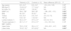
ResultsOut of 39 boys with DMD, 18 were excluded due to non-ambulatory status, and all the remaining ambulatory patients (n = 21) aged between 4 and 13 (8.00 ± 2.4 years) met the inclusion criteria and agreed to participate. Ten healthy boys aged between 6 and 13 (8.9 ± 2.4 years) were included as controls. Twenty of 21 patients’ genetic analysis results were available and a mutation was detected in 17 of 20 patients. Fourteen of 17 patients had distal hot-spot deletions encoding for exons between 44 and 55, three of them had proximal hot-spot deletions encoding for exons between 3 and 19. The clinical characteristics of the participants are summarized in Table 1. The boys with DMD had significantly worse scores in 6MWT, TUG, BBS, TRF, and MMT (both upper and lower limbs), and lumbar lordosis was significantly higher than the healthy controls. Age, BMI, fall index, and thoracic kyphosis were similar in both groups.
Clinical characteristics of the patients with DMD and the controls.
| Patients (n: 21) | Controls (n: 10) | Mean difference (95% CI) | P | |
|---|---|---|---|---|
| Age (years) | 8.0 ± 2.4* | 8.9 ± 2.4* | −0.9 (−2.9, 1.02) | 0.35† |
| BMI (kg/m2) | 15.8 (4.3)⁎⁎ | 17.7 (4.4)⁎⁎ | NA | 0.30‡ |
| fall index (%) | 100 (3)⁎⁎ | 100 (4)⁎⁎ | NA | 0.95‡ |
| 6MWT (m) | 364 ± 136* | 560 ± 65* | −196 (−291, −101) | 0.001† |
| BBS | 49 (12)⁎⁎ | 55 (0)⁎⁎ | NA | 0.001‡ |
| TUG(s) | 8.3 ± 4.4* | 3.9 ± 0.5* | 4.4 (1.5, 7.3) | 0.004† |
| TRF (s) | 3.8 (1.9)⁎⁎ | 1.9 (0.4)⁎⁎ | NA | 0.001‡ |
| Lumbar lordosis (°) | 34.4 ± 9.7* | 27.5 ± 4.7* | 6.9 (1.6, 12.1) | 0.01† |
| Thoracic kyphosis (°) | 25.7 ± 11.1* | 27.4 ± 3.7* | −1.6 (−10.5, 3.4) | 0.18† |
| MMT (%MRC) | ||||
| Upper limbs | 78.5 ± 12.6* | 95.4 ± 3.1* | −16.9 (−25.1, −8.7) | 0.001† |
| Lower limbs | 72.5 (11.2)⁎⁎ | 97.5 (6.5)⁎⁎ | NA | 0.001‡ |
6MWT, six minute walk test; BBS, Berg Balance Scale; BMI, body mass index; DMD, Duchenne Muscular Dystrophy; MMT, manual muscle test; MRC, Medical Research Council; NA, not applicable; TRF, time to rise from floor test; TUG: timed up and go test.
Bold data signify a statistically significant difference.
The results of repeated inclinometric measurements were statistically similar according to t-test (p = 0.58 for lumbar lordosis, 0.86 for thoracic kyphosis) and displayed excellent reliability (Cronbach's α = 0.95 for lumbar lordosis, 0.92 for thoracic kyphosis).
In the group with DMD, according to Vignos scale, seven boys were at level 1 (33%), seven boys were at level 2 (33%), four boys were at level 3 (19%), and three boys were at level 4 (14%). Twelve boys were wearing night splints (57%). Sixteen (76%) boys were on steroids, 12 of them were on daily, and four of them were on alternate days regimen. Five (24%) children had mild scoliosis with a mean Cobb angle of 12° ± 2.5° (min-max: 10°−15°). One of these boys had thoracic scoliosis (apex: T6), while the others had thoracolumbar scoliosis (apexes varied between T11 and L1).
There was a moderate positive correlation between lumbar lordosis and age (r = 0.532, p = 0.01), lumbar lordosis and BMI (r = 0.522, p = 0.01), and age and BMI (r = 0.633, p = 0.002). The amount of lumbar lordosis correlated strongly and negatively with 6MWT (r=−0.710, p = 0.001), and moderately and positively with TUG (r = 0.666, p = 0.002) and TRF (r = 0.462, p = 0.046), and moderately and negatively with BBS (r=−0.683, p = 0.05). The thoracic kyphosis did not correlate significantly with any of these variables (p > 0.05). There was a moderate and negative correlation between 6MWT and TUG (r=−0.565, p = 0.02), a lesser distance on the 6MWT being correlated with more time to perform the TUG (negative correlation due to worse performance being smaller value of 6MWT and bigger value on TUG). Upper limbs and lower limbs strength did not correlate with any of the variables (p > 0.05).
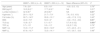
When the patients were stratified according to the distance covered in the 6MWT (below or above 350 m), the boys who could walk <350 m in 6 min had a significantly higher lumbar lordosis angle and worse scores in BBS and TUG. There were no significant differences in terms of age, BMI, fall index, kyphosis angle, TRF, and MMT between children who could walk less than or more than 350 m in 6 min (Table 2).
Comparison of the clinical characteristics of the patients with DMD after stratification according to the distance covered in the 6MWT.
| 6MWT < 350 m (n = 9) | 6MWT ≥ 350 m (n = 12) | Mean difference (95% CI) | P | |
|---|---|---|---|---|
| Age (years) | 7.9 ± 2.6* | 8.3 ± 1.6* | −0.3 (−2.5, 1.3) | 0.76† |
| BMI (kg/m2) | 18.5 (9.2)⁎⁎ | 17.7 (4.9)⁎⁎ | NA | 0.21‡ |
| Lumbar lordosis (°) | 42 (5.8)⁎⁎ | 28 (5.8)⁎⁎ | NA | 0.01‡ |
| Kyphosis (°) | 29.1 ± 12.3* | 21.7 ± 7.9* | 7.4 (−3.5, 10.0) | 0.22† |
| Fall index (%) | 90.7 ± 14.5* | 95.6 ± 10.1* | −4.9 (−17.8, 11.3) | 0.45† |
| BBS | 43.3 ± 7.6* | 53.0 ± 2* | −9.2 (−15.0, −6.8) | 0.01† |
| TUG (s) | 10.8 ± 3.7* | 5.1 ± 0.7* | 5.7 (4.4, 8.2) | 0.001† |
| TRF (s) | 3.7 ± 1.5* | 3.5 ± 1.3* | 0.2 (0.7, 3.5) | 0.79† |
| MMT -UL | 74.3 ± 11.4* | 80.0 ± 12.8* | −5.7 (−24.5, −3.1) | 0.37† |
| MMT-LL | 67.9 ± 10.7* | 72.5 ± 14.1* | −4.7 (−30.7, −3.9) | 0.48† |
6MWT, six minute walk test; BBS, Berg Balance Scale; BMI, body mass index; DMD, Duchenne Muscular Dystrophy; MMT-LL, manual muscle tests lower limbs; MMT-UL, manual muscle tests upper limbs; NA, not applicable; TRF, time to rise from floor test; TUG, timed up and go test.
aBold data signify a statistically significant difference.
Separate diagnostic performances of lumbar lordosis, BBS, and TUG tests were evaluated to assess their ability to discriminate the ambulatory function assessed with the 6MWT. According to the ROC analysis performed, AUC values were excellent for BBS and TUG (0.948, 0.906, respectively) and good for lumbar lordosis (0.850). Based on the maximum value of Youden Index, the optimal threshold was determined to be 36° for lumbar lordosis (Table 3).
Sensitivity, specificity, positive and negative likelihood ratio, positive and negative predictive values, and threshold value of lumbar lordosis for discriminating the 6MWT.
| AUC | P | Criterion | Sensitivity | Specificity | +LR | −LR | +PV | −PV | |
|---|---|---|---|---|---|---|---|---|---|
| lumbar lordosis | 0.85 | 0.003 | >36° | 87.5 | 90 | 8.7 | 0.14 | 87.5 | 90 |
6MWT, 6 min walk test; AUC, area under curve; LR, likelihood ratio; PV, predictive value.
aBold data signify a statistically significant difference.
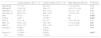
The boys whose lumbar lordosis was higher than 36° had significantly worse scores in BBS, TUG, and MMT of the upper and lower limbs and were more likely to have poorer functional levels. The amount of lumbar lordosis had no significant effects on the other variables (Table 4).
Comparison of the clinical characteristics of the patients after stratification according to the lumbar lordosis threshold.
| Lumbar lordosis < 36°(n = 11) | Lumbar lordosis ≥ 36°(n = 10) | Mean difference (95% CI) | P (%95 CI) | |
|---|---|---|---|---|
| Age (years) | 7.1 ± 2.1* | 9.0 ± 2.6* | −1.8 (−3.9, 0.3) | 0.09† |
| BMI (kg/m2) | 12.8 ± 0.4* | 16.6 ± 1.1* | −3.8 (−7. 2, −0.4) | 0.09† |
| Kyphosis (°) | 23.2 ± 5.9* | 23.4 ± 13.7* | 0.2 (−9.1, 10.7) | 0.86† |
| Fall index (%) | 95.6 ± 10.1* | 93.8 ± 12.3* | 1.8 (−9.5, 13.1) | 0.74† |
| BBS | 55 (1)⁎⁎ | 45 (18)⁎⁎ | NA | 0.004‡ |
| TUG (s) | 4.2(1)⁎⁎ | 10 (9.2)⁎⁎ | NA | 0.007‡ |
| TRF (s) | 3.7 ± 1.6* | 4.6 ± 2.7* | −0.9 (−3.8, 0.9) | 0.40† |
| MMT -UL | 84.8 ± 12.5* | 72.9 ± 9.6* | 11.9 (1.2, 22.6) | 0.03† |
| MMT-LL | 79.4 ± 13.7* | 65.0 ± 10.3* | 14.4 (7.7, 26.1) | 0.01† |
| Scoliosis + | 9 (56%) | 7 (44%) | 0.64# | |
| − | 2 (40%) | 3 (60%) | ||
| Vignos1–2 | 11 (78%) | 3 (22%) | 0.001# | |
| 3–4 | 0 (80%) | 7 (100%) |
BBS, Berg Balance Scale; BMI, body mass index; DMD, Duchenne Muscular Dystrophy; MMT-LL, manual muscle tests lower limbs; MMT-UL, manual muscle tests upper limbs; NA, not applicable; TRF, time to rise from floor test; TUG, timed up and go test.
aBold data signify a statistically significant difference.
The main result of this study is that ambulation and dynamic balance are negatively associated with the increment of lumbar lordosis with a cut-off point of 36° in boys with DMD. It is known that an increase in lumbar lordosis is a compensatory mechanism that occurs secondary to muscle weakness.23 However, our results suggest that balance and gait are affected by the increased lumbar lordosis before obvious strength loss occurs.
This study has several important limitations. The digital inclinometer, which was used to measure lumbar lordosis is reliable in healthy adults, but, although it has been used in pediatric studies,8,9 its reliability was not assessed among children. But, to address this limitation, we checked the intrarater reliability and found that it was very reliable as used in this study. Also, using a pediatric version of the BBS [Pediatric Balance Scale (PBS)] for balance evaluation would have helped us achieve more reliable results. However, a validity and reliability study of the PBS had not been performed for the language spoken by the patients on the date we conducted the study. Limitations related to muscle strength testing include lack of testing of the trunk musculature as well as the use of a method that is highly operator dependent. Normative values of sagittal spinal alignment of children are lacking in the literature, thus it is difficult to determine what values are normal. We have tried to overcome this issue by comparing boys with DMD with healthy control participants. As the functional tests were a part of a routine visit, the intrarater reliability of these tests in our setting has not been determined.
Muscle strength, which is a part of the efferent system of the central nervous system, is an important factor contributing to postural control and balance.24–26 An intact muscular system is required to maintain upright balance, during both stance and ambulation.25 Kids with DMD have poor pelvic control and have difficulty maintaining balance due to proximal weakness.25 However, MMT using the MRC is not a reliable tool to assess the strength of only mildly affected muscles.16 In children who are in an early stage of DMD, clinical or functional tests may not be sensitive enough to detect early compensatory strategies. Moreover, muscle strength has previously been found to be less sensitive than timed function tests to changes in disease status in ambulatory boys with DMD.10 McDonald et al.10 reported that, although there were significant declines in ambulatory function, relatively small changes occurred in muscle strength of patients with DMD. Our findings are consistent with these earlier findings because no significant relationship was detected between muscle strength and the 6MWT and balance tests. In addition, according to the current results, although lumbar lordosis angle was found to be increased in boys with DMD compared to controls, there seems to be no correlation between muscle strength and lumbar lordosis, which contradicts the knowledge that the increase in lumbar lordosis is caused by a decrease in muscle strength.2 These discrepancies may be explained by the muscle strength approaching the floor effect.
When a child with DMD becomes hyperlordotic due to weakness and/or shortness of pelvic girdle muscles, the center of mass demonstrates greater displacements which may lead to balance and gait disturbances.2 Similarly, in the current study, the increase in lumbar lordosis was found to be highly correlated with the ambulation function measured by the 6MWT. Also, significant relationships were detected between lumber lordosis and dynamic balance tests in terms of TUG and TRF, consistent with previous studies.1–3 The main question of the present study was at what degree of lumbar lordosis these alterations start. Based on the ROC analysis, 36° has been determined as a threshold of gait deterioration. And the patients with lumbar lordosis over 36° were shown to have worse scores in dynamic balance tests, muscle strength, and functional level.
Horlings et al.25 and Kaya et al.26 investigated the effects of muscle weakness on balance and found that patients with DMD with proximal weakness had worse scores in static balance than the distal weakness group. Our data showed that dynamic balance was better correlated with ambulation and disease severity than static balance evaluated by posturography. It should be noted, however, that all the patients in this study were at high-risk category according to posturography (median fall index: 100), which might have resulted in a ceiling effect. But, although Kohen-Raz15 reported that the reliability for posturography scores collected among 43 healthy children in second and third grades was between 0.68 and 0.95, because this test was not capable of discriminating between kids with DMD and healthy kids in the present study, a suspicion arises about the validity of the test among children. So, we cannot simply claim that static balance has no relationship with sagittal alignment or functional performance.
ConclusionAn increase in lumbar lordosis has negative effects, especially on dynamic balance and ambulation, in children with DMD. Therefore, to maintain balance and ambulation, it is recommended to monitor these patients in terms of sagittal curvatures such as lumbar lordosis, and tailor physical therapy programs accordingly.
The authors thank all the children and their families for participating in this study. This research was supported by Health Sciences University, Antalya Training and Research Hospital without any involvement in the study design, in the collection, analysis and interpretation and of data; in the writing of the manuscript; or in the decision to submit the manuscript for publication.


