


Runners seek health benefits and performance improvement. However, fatigue might be considered a limiting factor. Transcranial Direct Current Stimulation (tDCS) has been investigated to improve performance and reduce fatigue in athletes. While some studies showing that tDCS may improve a variety of physical measures, other studies failed to show any benefit.
ObjectiveTo evaluate the acute effects of tDCS on central and peripheral fatigue compared to a sham intervention in recreational runners.
MethodsThis is a triple-blind, controlled, crossover study of 30 recreational runners who were randomized to receive one of the two interventions, anodal or sham tDCS, after the fatigue protocol. The interventions were applied to the quadriceps muscle hotspot for 20 min. Peak torque, motor-evoked potential, and perceived exertion rate were assessed before and after the interventions, and blood lactate level was assessed before, during, and after the interventions. A generalized estimated equation was used to analyze the peak torque, motor-evoked potential, and blood lactate data, and the Wilcoxon test was used for perceived exertion rate data.
ResultsOur findings showed no difference between anodal tDCS and sham tDCS on peak torque, motor-evoked potential, blood lactate, and perceived exertion rate.
ConclusionThe tDCS protocol was not effective in improving performance and reducing fatigue compared to a sham control intervention.
Brazilian Clinical Trials RegistryRBR-8zpnxz.
Runners are constantly searching for ways to improve their performance. However, fatigue can be a limiting factor, as it can cause a reduction in tension, strength, and muscle contraction speed.1,2 Furthermore, once present, fatigue alters cadence, stride length, and lower limb joint kinematics during running,3–5 which may lead to poor performance. Fatigue is defined as any reduction in the ability to produce maximum muscle strength after prolonged physical activity,3 and may be classified as having central or peripheral origin.5 In central fatigue, the reduction in muscles’ voluntary activation may be directly related to the decreased frequency and synchronization of motor neurons due to changes in the neurotransmitter levels, which leads to neural impulse failure from the motor cortex to the motor neurons.6,7 In peripheral fatigue, there is a decrease in the muscle fibers’ contractile strength, with changes in extracellular metabolism.3,8
To maintain strength during the muscle fatigue process, motor neuron inputs in the motor cortex should increase. The increase in motor cortex excitability can enhance the downward drive, resulting in sustained neural activation from motor neurons to active muscles, thereby improving muscle output and delaying the onset of fatigue. This neuronal excitability can be elicited through Transcranial Direct Current Stimulation (tDCS).9 Sasada et al10 reported that tDCS may be a promising technique in central fatigue, as it can improve neural drive excitability and delay fatigue onset.
In the sports field, previous studies investigating the effects of tDCS on performance and fatigue showed improvements in exercise tolerance11 and jumping performance in men with experience in strength training12 and parkour athletes.13 In contrast, there is evidence showing no beneficial effects of tDCS on performance.14 In addition, Davis15 argues that the use of these technologies for enhancing athletes’ performance during exercise training should not be considered unethical in sports. Nonetheless, given the contradictory results, the usefulness of tDCS remains warranted.
Therefore, this study investigated the role of tDCS as a recovery strategy to reduce muscle fatigue in athletes. More specifically, we investigated the acute effects of tDCS, performed before a fatigue protocol, on quadriceps muscle strength (i.e. peak torque) compared to a sham tDCS in recreational runners. We also investigated the effects on cortical excitability, blood lactate level, and rate of perceived exertion (RPE).
MethodsTrial designIt is a triple-blind, sham-controlled crossover clinical trial following the protocol published by Uehara et al.16 This study follows the Consolidated Standards of Reporting Trials (CONSORT) statement. This trial was approved by the Research Ethics Committee of the Nove de Julho University (CAAE certificate: 26427819.1.0000.5511) and was registered in the Brazilian Clinical Trials Registry (ReBEC) (RBR-8zpnxz). All participants signed an informed consent document before participating in the study.
ParticipantsParticipants were invited to participate in the study by advertisements through social networks and contacting recreational runner groups in the region where the study was conducted. Adult recreational runners who ran from 5 to 21 kilometers, three times a week, for at least six months were considered eligible for this study. Pregnant women, people who practiced other sports, who had recent musculoskeletal injuries, recurrent seizure history, skin lesions on the stimulation area, who used antihistamines, antidepressants, antiepileptic, pacemakers, or who had metal implants in the head were excluded from the study. Participants were instructed not to modify their training during the study.
InterventionsParticipants attended the research laboratory three times, with an interval of seven days between each session. In the first session, we collected the participants’ demographic data. In the second and third sessions, participants received the intervention protocol consisting of the anodal tDCS or the sham tDCS followed by the fatigue protocol, according to the randomization schedule (Fig. 1). Participants were instructed to avoid consuming caffeine, tea, alcohol, tobacco, and drugs and not perform physical activities the day before interventions.

Participants were classified as responders or non-responders to the tDCS before the protocol. Motor-evoked potential (MEP) was measured immediately before and after anodal tDCS stimulation at 2 mA for five minutes. Then, those participants with initial and final MEP ratios greater than 1.0 were considered responders.17
The anodal tDCS was applied before the fatigue protocol, using the NeuroConn DC-STIMULATOR (Germany) for 20 min, 2 mA intensity, 30-s linear ramp up/down, using two non-metallic surface electrodes, 5 × 7 cm (cathode) and 5 × 5 cm (anode). The anode electrode was positioned over the dominant quadriceps muscle hotspot, previously established using TMS.16 The cathode was placed on the supraorbital contralateral region. For the sham tDCS, the same set up used for anodal tDCS was applied to the sham tDCS, except that the equipment was turned off after 20 s.
Fatigue protocolThe fatigue protocol follows the methods by Schwendner et al.18 The participants were placed in an isokinetic dynamometer (Biodex System 2), having a 100° angle between the trunk and hip, and the lower dominant member at a 60° flexion (0° corresponding to full knee extension). The dynamometer axis was placed at the knee´s rotational axis.
The participants executed flexion and extension concentric contractions of the dominant limb knee, at 100° of maximum voluntary contraction (MVC), at a 60°/s speed , with a 60° range of motion (between 90° and 30° knee flexion). For each contraction, there was an opposing force provided by the dynamometer. Muscular fatigue was considered when the participant did three contractions at less than 50% of MVC (pre-established in isokinetic dynamometer).
MeasurementsPeripheral fatigue was assessed by collecting information on the following variables: peak torque, blood lactate level, and RPE. Central fatigue was determined by assessing the cortical excitability via MEP. The peak torque, MEP, and RPE were performed before and after the intervention protocol. Blood lactate was evaluated before, during, and after the fatigue protocol.
Primary outcomePeak torque was the primary outcome. To assess peak torque, the participant performed three isometric quadriceps contractions of the dominant limb in an isokinetic dynamometer (Biodex System 2) for 10 seconds for each contraction. The highest contraction torque value out of the three attempts was considered for the analysis.
Secondary outcomesSecondary outcomes of this study were MEP, blood lactate level, and RPE. The MEP was assessed by the MagPro R30 (Magventure, Denmark) Transcranial Magnetic Stimulation (TMS), with a 70 mm diameter figure-of-eight coil, using biphasic pulses and MepOption (Megaventure, Denmark) to capture the myoelectric signals. The coil was positioned over a motor area (M1) to locate the quadriceps hotspot. To capture the myoelectric signal disposable silver chloride electrodes were placed on the rectus femoris ventral surface muscle with a 2 cm inter electrode distance, and the reference electrode was set up on the C7 vertebra spinous process. These positions followed the Surface Electromyography for Non-Invasive Muscle Assessment protocol.
The resting motor threshold (rMT) was estimated considering the lowest TMS intensity to generate an MEP >50 µV peak-to-peak amplitude. Twenty-four pulses at 120% of rMT were employed to measure MEP from quadriceps hotspot before and after fatigue protocol to evaluate cortical excitability.19 The participants who did not take an MEP >50µV peak-to-peak amplitude were excluded from the analysis.
For the analysis of blood lactate level, 25 µl blood samples were collected through the index finger puncture, with lancets (Nanolet™). Blood collection was performed before (lactate 1), during (lactate 2), and two minutes after the end of the fatigue protocol (lactate 3). Lactate levels were compared between the anodal and sham tDCS for each time point.
The RPE was assessed using the modified Borg Scale20 during the muscular fatigue protocol. RPE scores ranged from 0 (no effort) to 10 (extremely difficult).
Side effectsThe potential side effects of tDCS were investigated by applying the tDCS Side Effects Questionnaire21 after each intervention, containing questions such as: Did you have any of these sensations (pain, fatigue, burning sensation, itching, heat, others)? Data were expressed as absolute frequency distribution for each group.
Sample sizeThe sample size was calculated using the GPOWER3 software. The paired t-student test was used considering the difference in peak torque results between post and pre-tDCS intervention of five participants in the anodal tDCS (44.66±15.25 N.m) and sham tDCS (24.96±10.64 N.m). Based on the difference between groups, using a power of 90% and an alpha level set at 0.05, 16 participants were required. However, 30 participants were recruited to allow for possible dropouts.
RandomizationParticipants were randomized to receive the anodal tDCS or the sham tDCS in the first session. In the second session, participants received the other intervention. An investigator not involved in providing treatments or assessing participants was responsible for the randomization process. Randomization was performed using the website www.randomizer.org.
BlindingThis study was triple-blind (assessors, therapists, and participants). Assessor blinding was possible because the assessors who measured the outcomes were blinded to the treatment received by each participant. The NeuroConn DC-STIMULATOR device has a configuration in which, through codes, the active or simulated mode is selected, so the researcher, who conducted the treatment, and the patient, did not know which mode was chosen. To validate the blinding of participants,22 participants answered at the end of each intervention the following question: “What type of treatment did you receive, the sham or the active tDCS?”.
Statistical analysisStatistical analyses were performed using the SPSS (IBM; v.25.0). Only participants who completed both intervention sessions were considered for analysis. Data on peak torque, MEP, blood lactate level, and RPE were analyzed considering within-group change and between-group comparison. Data were presented as estimated marginal means and 95% confidence interval (CI). The statistical test used was Generalized Estimated Equations followed by Bonferroni post-hoc test for multiple comparison adjustments. For peak torque and MEP amplitude, two fixed factors were used: group [anodal tDCS, sham tDCS] x time [pretest, posttest]; for blood lactate two fixed factors were used: group [anodal tDCS, sham tDCS] x time [pre, during, post fatigue protocol]; for RPE the Wilcoxon test was used. Statistical significance was set at p < 0.05.
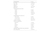
ResultsThirty healthy recreational runners (39.1 ± 6.8 years) participated in the study. Their clinical demographic characteristics are described in Table 1. Seventy percent of participants practiced physical activity more than five times a week; 60% had a good or excellent cardiorespiratory capacity, and around 66% had good sleep quality.
Descriptive characteristics of the sample (N = 30).
Data are mean (standard deviation) or frequency (proportion).
BMI, Body Mass Index; VO2Max, Maximal Oxygen Consumption, tDCS: transcranial direct current stimulation.
Data for primary and secondary outcomes are summarized in Table 2. The peak torque at baseline was similar between groups. The within-group change analysis showed a significant decrease in peak torque after the fatigue protocol in both groups. For the between-group comparison, the anodal tDCS group showed a similar reduction in peak torque of the quadriceps muscle compared to the sham tDCS group.
Primary and secondary outcomes comparisons before and after intervention for the study groups.
| Anodal tDCS (N = 30) | Sham tDCS (N = 30) | Between-group differencea | |
|---|---|---|---|
| Peak torque (N.m) | −5.5 (−19.9, 9.0) | ||
| Baselinea | 207.5 (186.6, 228.4) | 205.1 (183.0, 227.1) | |
| Post-interventiona | 189.7 (171.8, 207.7) | 192.8 (176.6, 209.0) | |
| Within-group changea | −17.8 (−33.0, −2.5) | −12.3 (−26.3, 1.7) | |
| MEP amplitude (µV) | −51.8 (−141.9, 38.3) | ||
| Baselinea | 272.2 (222.5, 321.9) | 241.9 (192.5, 291.2) | |
| Post-interventiona | 185.0 (114.8, 255.3) | 197.6 (139.3, 255.9) | |
| Within-group changea | −87.1 (−189.7, 15.4) | −44.3 (−137.1, 48.5) | |
| Lactate (mmol•l−1) | |||
| Baselinea | 2.3 (2.0, 2.7) | 1.9 (1.4, 2.4) | −0.7 (−0.5, 1.9) |
| During the protocola | 5.8 (4.6, 7.1) | 4.7 (3.6, 5.8) | |
| Change during the protocol minus baselineb | 3.5 (1.9, 5.1)* | 2.3 (0.7, 4.0)* | |
| Post-interventiona | 5.8 (4.7, 6.9) | 5.3 (4.2, 6.4) | −0.7 (−1.7,0.4) |
| Change post-intervention minus during the protocola | −0.0 (−0.9, 0.9) | 0.6 (−0.6, 1.9) | |
| RPE (rating)b | 7.7 [7, 9] | 7.6 [7, 8.3] |
For the MEP analysis, six participants from the anodal tDCS (n = 24) and five from the sham tDCS (n = 25) were not included in the analysis because the MEP peak-to-peak amplitude was less than 50µV. Our results for the within-group change revealed no significant reduction in MEP in both groups. In addition, no difference between the anodal and sham tDCS groups was found (Table 2).
The blood lactate levels were similar for both groups at baseline. The within-group changes in the lactate levels were significant from baseline to during the protocol but non-significant from during the protocol to post-intervention in both groups. No difference in blood lactate levels was found between the groups during the protocol and post-intervention (Table 2). RPE was assessed during the fatigue protocol. Participants in both groups showed similar perceived exertion rates.
For side effects, the anodal tDCS group reported itching (n = 1), pain (n = 1), burning sensation (n = 2), pinching (n = 2), metallic taste (n = 1), and fatigue (n = 3). Similarly, the sham tDCS group reported itching (n = 1), pain (n = 2), burning sensation (n = 4), heat (n = 4), pinching (n = 2), and fatigue (n = 2). Rates of side effects were similar in both groups.
The assessment of blinding showed that only 16.66% of the participants (3 participants receiving sham tDCS and 2 participants receiving anodal tDCS group) could identify which stimulation, active or sham placebo, they received.
DiscussionOur findings showed that a session of tDCS on the M1 before a protocol of quadriceps muscle fatigue did not reduce fatigue in adult recreational runners. Comparison between groups showed no difference between anodal tDCS and sham tDCS.
The results of studies that used tDCS on M1 are diverse, as Codella et al23,24 observed an increase in the power and resistance of the lower limbs during running, while Giboin & Gruber25 observed that both cathodal tDCS and anodal tDCS impaired force production during an intermittent fatigue maximal voluntary contraction task. A study by Machado et al26 observed a delay in central fatigue by stimulation in M1 and, Chen et al27 observed an improvement in the performance of repeated sprints after anodal tDCS. Our results contradicts some of these previous studies on the effects of anodal tDCS on athletes' performance. The effects for each of the outcome variables are discussed separately below.
Peripheral fatigueThe reduction in peak torque after the fatigue protocol was predictable because physiological changes occur after fatiguing tasks, for example, changes in pH and cellular metabolites. These changes lead to a reduction in contractility and muscle strength.8,28 Our study hypothesis was that anodal tDCS applied to the quadriceps hotspot before the fatigue protocol would minimize fatigue (i.e., less reduction in peak torque for the tDCS group anodic); however, this hypothesis was not confirmed. However, Codella et al23 observed an increase in power and endurance of the lower limbs during running in healthy physically active men after tDCS, and Park et al24 observed an increase in exercise duration after tDCS was applied during running up to exhaustion. Other studies11,29,30 also demonstrated a reduction in fatigue assessed through time to exhaustion and motor failure tests; however, they were performed on healthy, physically active participants.
These results may have occurred because tDCS can modulate cortical excitability, increasing the neuronal drive of the locomotor muscles located in M1.29 However, for this effect, more than a single session of tDCS may be necessary, as reported by some studies31–34 which indicates that a single session of tDCS in M1 failed to improve maximal anaerobic exercise. Giboin and Gruber25 also did not observe the effects of tDCS on knee extensor strength applied during and before the fatiguing task. This result may occur because under conditions of maximum strength, the muscles are already working at maximum, so all motor units are already recruited; therefore, ceiling effects do not allow tDCS to show additional effects.12,13
Another factor to be considered is that we used a 2 mA intensity, as in other studies with tDCS and fatigue.11,25,29 However, Workman et al35 observed better effects on peak torque after the fatigue protocol in physically active adults when tDCS was applied at 4 mA compared to 2 mA. Despite this, few studies investigate the stimulation's effects with intensities greater than 2 mA. Therefore, further studies with higher intensities should confirm whether stimulation intensity could interfere with tDCS’ effects on fatigue.
There was a significant increase in lactate 2 and 3 compared to lactate 1 levels in both groups, which was expected, because lactic acid tends to increase proportionally to exercise intensity, being considered a fatigue marker36,37 However, as there was no difference between the groups, the anodal tDCS did not interfere with the lactate level. The same was observed in elite swimmers who received tDCS.38
The RPE results of the groups in our study increased, without significant differences between them. Similarly, Baldari et al39 did not observe the tDCS’ effects on the RPE of recreational runners during the incremental ramp test. However, Williams et al30 observed improvements in RPE in healthy participants who received anodal tDCS during elbow flexors submaximal sustained contraction. The difference in results may be because previous studies evaluated smaller muscle groups; therefore, the exertion feeling would seem more easily recoverable.
Central fatigueThe MEP results show a reduction in both groups, which is expected after fatiguing tasks because there is a decrease in the motor neurons firing due to the excitability depletion or in the excitatory synapse response.5
However, we hypothesized that anodal tDCS applied over the quadriceps muscle hotspot could reduce fatigue compared to the sham tDCS, but we did not find these results. In contrast, Angius et al33 observed in active men an increase in MEP amplitude after anodal tDCS in the knee extensors isometric exhaustion test, and Kristiansen et al40 observed an increase in cortical excitability after anodal tDCS in physically active individuals, but without change on fatigue time on bike and RPE.
We argue that our findings regarding MEP are due to individual variability, as our participants were not homogeneous concerning functional capacity and kilometers covered in the race. Furthermore, of the 30 participants, only seven were tDCS responders. Therefore, repeating this protocol with more participants responsive to the stimulation technique may produce different results. Another hypothesis is that in a longitudinal study, we may find different results, as observed by Lopez-Alonso et al,17 who evaluated intra- and inter-subject variability in healthy individuals and observed a 44% increase in responders to tDCS after the second session compared to the first session. In addition, the literature shows us that the tDCS’ effects are summative and stimulate neuroplasticity.41 This effect was demonstrated by Ammann et al42 who reported an increase in the healthy individuals’ MEP amplitude from the first to the third tDCS session; moreover, longitudinal evidence of performance improvement through the M1 tDCS was provided in adolescents’ professional rowing athletes.43
However, our view is that further research is still needed to investigate whether tDCS is for everyone. Esteves et al44 report that the training level can affect neuronal activity and brain structure, being able to produce different responses to stimulation. It has been shown that tDCS has more effect when individuals have a lower baseline activity or experience, i.e., when changes in synaptic connectivity are required; therefore, when there is the possibility of neuron functional improvements. Despite being recreational runners, almost all participants in the current study practiced exercises more than five times a week, which may have altered the motor threshold and cortical excitability, making it laborious to obtain a response. Performance improvement becomes increasingly arduous to achieve as the performance level rises due to the so-called “ceiling effects”.45
A recent review46 that included 19 studies looked at the tDCS acute effects on changes in athletes' motor performance compared to simulation and showed that anodal tDCS led to better performance in athletes on sport-specific motor tasks, but none of the athletes in the review were recreational runners. Of the 19 studies included, 10 investigated resistance effects; five found an increase in specific endurance performance, while five showed nothing.
Therefore, we believe that, for clinical practice, more studies are necessary to evaluate the participants' neurophysiological functions who received more than one intervention session, considering the variability that can interfere with the results, such as age, sex, skull, and brain shape.47 Thus, individualized stimulation protocols are necessary to understand how tDCS works for athletes. As with other therapies and medications, for example.
Finally, a last hypothesis for not finding a tDCS effect would be the rise in cortisol and glycogen levels. Although we did not perform this measurement, Mellow et al48 state that after high-intensity exercises, as the fatigue protocol, there is an increase in the cortisol and glycogen levels that can block the non-invasive stimulation effects on neuroplasticity. Thus, these measures may be relevant for future studies involving tDCS and fatigue.
Limitations of this study and future perspectivesAs limiting factors for this study, we can highlight the participants’ heterogeneity concerning the distance they run, as we had eight 5 km runners, eleven 10 km runners, and eleven 21 km runners. Furthermore, they presented different life and training history. López-Alonso et al17 reported this same difficulty, noting a large inter-individual variability in the response to tDCS, explaining the difference in results.
Of the 30 participants, only seven were tDCS responders. We believe that this study reinforces the individual´s evaluation importance and that future studies evaluate the longitudinal tDCS effects within the specific functions of each sport.
However, it is necessary to note that brain stimulation may be considered as a “neuro-doping” agent, as it can improve physical and mental performance in sports. Therefore, with the growing field of brain stimulation for performance, this question may be one relevant point for debate.
ConclusionWe conclude that the tDCS session on the quadriceps cortical motor area applied before the fatigue protocol does not affect the central and peripheral fatigue in recreational runners.
We thank the foundation for Coordenação de Aperfeiçoamento de Nível Superior (Coordination for the Improvement of Higher Education Personnel, CAPES, acronym in Portuguese), Conselho Nacional de Desenvolvimento Científico e tecnológico (the National Council for Scientific and Technological Development, CNPq, acronym in Portuguese), and the Nove de Julho University for the support they provided. Those providing financing had no role in the design of this study, data collection, and analysis, decision to publish, or preparation of the manuscript. We also thank the Transcranial Electrical Stimulation Laboratory (TESLAB) and Correlatos Neurais do equilíbrio e da marcha (Neural Correlates of Balance and Gait, CONEM, acronym in Portuguese) of the Federal University of ABC for their partnership.


