


Airway clearance techniques include positive expiratory pressure, commonly used in our clinical practice, and a recently introduced temporary positive expiratory pressure device called UNIKO®. It is unclear which one provides the best benefit to patients.
ObjectivesThe aim of this observational 4-year study was to retrospectively compare the efficacy of and specific indications for temporary positive expiratory pressure compared to positive expiratory pressure in a standard rehabilitation program.
MethodWe retrospectively collected data from 162 subjects (107 males, mean age 70±9 years, 97 with primary diagnosis of chronic obstructive pulmonary disease, 65 with bronchiectasis), 51 treated with temporary positive expiratory pressure and 111 with positive expiratory pressure.
ResultsSubjects showed significant improvement in ratio of partial pressure arterial oxygen and fraction of inspired oxygen (p<0.001), forced vital capacity, forced expiratory volume in one second, peak expiratory flow, arterial oxygen saturation, and partial pressure arterial oxygen with no significant difference between positive expiratory pressure and temporary positive expiratory pressure groups apart from forced expiratory flow, which increased only in the positive expiratory pressure group. Evaluating specific subgroups, temporary positive expiratory pressure was more effective than positive expiratory pressure in improving gas transfer in subjects with emphysema and in those on oxygen therapy, as the effective supplement oxygen flow decreased significantly (p=0.034 and 0.046 respectively for temporary positive expiratory pressure vs. positive expiratory pressure). In subjects on mechanical ventilation, positive expiratory pressure was superior to temporary positive expiratory pressure in increasing forced expiratory flow (p=0.018).
ConclusionThe physiological parameters of both groups improved significantly and similarly. Subgroup analysis suggests that temporary positive expiratory pressure could provide some advantage to subjects with emphysema and those on oxygen therapy, while positive expiratory pressure would benefit patients on mechanical ventilation. Randomized clinical trials are necessary to confirm our preliminary results indicating that different subgroups/phenotypes can benefit more from one type of treatment.
Patients with chronic hypersecretion (CH) of tracheobronchial mucus represent a challenging clinical problem. CH is a socially disabling condition causing a high susceptibility to airway infection and frequent, difficult-to-treat acute exacerbations with a significant impact on prognosis, quality of life, and use of health care resources, particularly in subjects with severely impaired respiratory function and/or chronic respiratory failure (CRF).1–5
Airway clearance techniques (ACTs), commonly performed by respiratory physiotherapists, are intended to aid secretion mobilization and expectoration and to mitigate complications associated with secretion retention.6 There is a lack of evidence on the superiority of any particular ACT or device and the guidelines do not support their routine use in these subjects,6 most of whom have chronic bronchitis (CB), chronic obstructive pulmonary disease (COPD), and/or bronchiectasis, even during an acute exacerbation.7 Nevertheless, ACTs still remain a challenging option of pulmonary rehabilitation (PR) programs.8,9
ACTs consist of a variety of approaches, such as forced exhalation, manual compression, and/or vibration of the thorax, and positive expiratory pressure (PEP) breathing.10,11 Three main clinical indications for PEP are described: to increase lung volumes, i.e., functional residual capacity (FRC) and tidal volume (VT), to reduce hyperinflation, and to improve airway clearance.12 One of the best-known methods to generate increased PEP is to breathe through a resistance device, such as a PEP mask.
More recently, the mechanical generation of low-level PEP during the main part of expiration (temporary positive expiratory pressure, TPEP) has been proposed to enhance airway clearance. A recent randomized clinical trial comparing the addition of TPEP to usual care13 showed an improvement in the level of dyspnea and of some physiological parameters in the TPEP group.
The aim of this retrospective study was to analyze data from our clinical experience over a 4-year period involving ACTs as part of a comprehensive PR program, in order to compare the physiological outcomes (i.e., gas exchanges and lung volumes) of two groups of patients treated with PEP mask and TPEP respectively. We also sought to identify specific indications for different ACTs in different subpopulations of subjects with CH.
MethodStudy design and subjectsWe chose to analyze data collected over 4 years from 162 CH patients admitted to the Department of Pulmonary Rehabilitation in the Salvatore Maugeri Foundation Institute of Veruno (NO), Italy, with primary diagnosis of CB/COPD or bronchiectasis, since these conditions are characterized by symptoms (such as chronic and progressive dyspnea, cough, and augmented sputum production14) and on the basis of the presence of CH as clinically defined by an abundant volume of daily sputum (>30mL/die).15 All patients had clinical signs and symptoms of CH, e.g., a history of frequent exacerbations, hospitalizations, and frequent use of antibiotics. On admission, subjects consented to the use of their clinical data for scientific purposes. The study was approved by the Internal Review Board at the Veruno Medical Center, Veruno (NO), Italy.
At the time of admission, CB was defined by the presence of cough and sputum for at least 3 months of the year for two consecutive years. COPD patients were staged and treated according to Global Initiative for Chronic Obstructive Lung Diseases (GOLD) criteria.14 Diagnoses of bronchiectasis and emphysema were confirmed by high-resolution computed tomography (HRCT) of the chest.16 Subjects with severe and/or unstable comorbidities (i.e., severe concomitant cardiovascular or neoplastic disease), which might have limited or impeded the execution of ACTs, were excluded from analysis.
Subjects were classified as being on oxygen therapy (OT) if on continuous oxygen treatment (24h) or only during sleep and/or exercise. They were classified as being on nocturnal positive pressure mechanical ventilation (MV) if prescribed by the attending physician. COPD exacerbation and bronchiectasis exacerbation were defined according to existing guidelines.14,15,17 In addition, subjects included in this analysis underwent ACTs with PEP-mask or TPEP (Fig. 1).
Treatments
Subjects were selected and underwent a comprehensive PR program in accordance with the existing ATS/ERS Joint Statement on Pulmonary Rehabilitation,1 individually tailored and designed, taking into account disabilities and tolerance to exercise. All patients performed two daily training sessions on a cycle ergometer or treadmill and unsupported upper limb exercises, graded as the subject progressed in the program. Subjects with CRF were provided with ambulatory oxygen as needed during training sessions. The need for ACTs was defined by the physiotherapists, in agreement with the physician in charge, based on symptoms and signs of excessive and/or retained secretions.
TPEP was approved in 2005 by the Ministry of Health as a therapeutic device for airway clearance. In 2006, the physical therapists of our Department were adequately trained to deliver TPEP treatment as an alternative to PEP-mask. At that time, they were not aware of the subsequent collection of data, so allocation to PEP-mask or TPEP treatment was based on patient preference, including comfort dyspnea and collaboration during the first ACT trial. Based on a retrospective analysis of the data collected between 2006 and 2009, we excluded patients with severe or unstable diseases, without CB/COPD or bronchiectasis, not treated with ACTs, or missing pre- or post-treatment. A total of 162 patients with CH using PEP (n=111) or TPEP (n=51) were included for analysis (Fig. 1).
Patients using PEP-mask performed two 15-min daily cycles. They were instructed to reach and maintain the highest mid-expiratory pressure tolerated between 10 and 20cmH2O (fixed by a manometer weekly) breathing at slightly increased VT, but not to use force at the end of the expiration. About every 2min, they performed a forced expiratory technique (FET) maneuver, huffing, and/or coughing without the resistor.12
Patients using TPEP performed two 15-min daily treatments with UNIKO (Medical Products Research, Legnano, Italy). They were asked to blow through a mouthpiece keeping TPEP active as long as possible for every breath and to cough as needed or at least every 3–5min. Both techniques were administered in the sitting position with elbows resting on a hard surface in front of them. Side effects or adverse events associated with treatments were recorded by the physiotherapists in charge.
Only data from subjects who underwent ACTs for at least 10 days within the in-hospital rehabilitation period were considered. ACTs were stopped at the end of PR program, corresponding to discharge from the Department.
Outcome parametersPhysiological parameters, i.e., data from spirometry and arterial blood gas analysis, measured both at baseline and at discharge were considered as primary endpoints based on previous data on the treatment of CH in severe patients.18 Dyspnea, other symptoms, and characteristics of secretions were not collected in a standardized way at that time and thus were not considered in our analysis.
Respiratory function was assessed using standard spirometry (6200 Auto box Pulmonary Function Laboratory, Sensormedics, Yorba Linda, CA, USA). Results were expressed as absolute values and percentage of their predicted values when indicated. Arterial oxygen saturation (SaO2) was monitored during treatments for safety procedures.
Arterial blood gases (ABGs) were determined in a radial artery blood sample, with the patient in resting condition, breathing room air or oxygen at the prescribed flow rate (fraction of inspired oxygen – FiO2). Both spirometry and ABG analysis (ABL analyzer, Radiometer, Copenhagen, DK) were performed according to existing guidelines.19
Statistical analysisDescriptive statistics were calculated for all variables; means and standard deviations or frequencies and percentages were reported according to the quantitative or qualitative nature of the variables, respectively. Baseline characteristics were compared using unpaired Student's t test or chi-square test, as appropriate. Repeated measures analysis of variance with one factor (TPEP vs. PEP) was used to test differences between pre- and post-assessments in the TPEP and PEP groups. A statistically significant interaction term was interpreted as a significant difference in pre- and post-effect between two techniques. The same analysis was applied to assess differences in subgroups identified according to clinically relevant variables (emphysema, OT, MV).
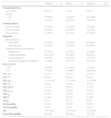
ResultsSubject characteristicsThe baseline demographic, clinical, and physiological characteristics of all subjects and comparison between groups (TPEP vs. PEP mask) are shown in Table 1. Most of them were male (66%), mean age 70 years (range 35–89), and current or former smokers; 60% had a primary diagnosis of CB/COPD and 40% had bronchiectasis.
Characteristics of the studied population.
| T-PEP (n=51) | PEP (n=111) | TOTAL (n=162) | |
|---|---|---|---|
| Demographic data | |||
| Age (years) | 68 (11) | 71 (9) | 70 (9) |
| Gender | |||
| M | 35 (69%) | 72 (65%) | 107 (66%) |
| F | 16 (31%) | 39 (35%) | 55 (34%) |
| Smoking history | |||
| Current smoker | 12 (24%) | 21 (19%) | 33 (20%) |
| Former smoker | 24 (47%) | 52 (47%) | 76 (47%) |
| Never smoked | 15 (29%) | 38 (34%) | 53 (33%) |
| Diagnosis | |||
| Main diagnosis | |||
| CB/COPD | 26 (51%) | 71 (64%) | 97 (60%) |
| Bronchiectasis | 25 (49%) | 40 (36%) | 65 (40%) |
| Additional diagnoses/conditions | |||
| Emphysema | 22 (43%) | 43 (39%) | 65 (40%) |
| Acute exacerbation | 26 (51%)* | 38 (34%)* | 64 (39%) |
| Oxygen therapya | 27 (53%) | 49 (44%) | 76 (47%) |
| Nocturnal mechanical ventilationa | 11 (22%) | 19 (17%) | 30 (19%) |
| Days of ACT | |||
| <15b | 18 (35%) | 35 (32%) | 53 (33%) |
| >15 | 33 (65%) | 76 (68%) | 109 (67%) |
| FVC (L) | 2.1 (0.7) | 2.0 (0.7) | 2.0 (0.7) |
| FVC% | 69 (22) | 69 (21) | 69 (21) |
| FEV1(L) | 1.15 (0.54) | 1.13 (0.47) | 1.14 (0.49) |
| FEV1% | 49 (23) | 52 (24) | 51 (23) |
| FEV1/FVC | 55 (14) | 58 (14) | 57 (14) |
| FEF25–75% | 22 (20) | 26 (22) | 25 (21) |
| FEF50% | 22 (21) | 25 (23) | 24 (22) |
| PEF% | 52 (22) | 50 (19) | 51 (20) |
| SaO2% | 93 (4) | 93 (3) | 93 (4) |
| PaO2(mmHg) | 66 (14) | 66 (10) | 66 (12) |
| PaCO2(mmHg) | 43 (9) | 43 (9) | 43 (9) |
| pH | 7.42 (0.03) | 7.42 (0.03) | 7.42 (0.03) |
| PaO2/FiO2(mmHg) | 282 (76) | 294 (65) | 291 (70) |
Data expressed as mean (SD) or n (%). COPD, chronic obstructive pulmonary disease; OT, oxygen therapy; MV, nocturnal positive pressure mechanical ventilation; FEV1, forced expiratory volume in one second % predicted; FVC, forced vital capacity % predicted; FEF25–75% and FEF50%, forced expiratory flows at 25–75% and at 50% of FEV1; PEF, peak expiratory flow % predicted; SaO2, arterial oxygen saturation; PaO2, partial pressure arterial oxygen; PaCO2, partial pressure arterial carbon dioxide; PaO2/FiO2, ratio of partial pressure arterial oxygen and fraction of inspired oxygen.
The sample was composed of subjects with moderate to severe lung disease. A large proportion (92/162) of them were CRF requiring OT and/or MV. Subjects showed a wide range of lung function values. Overall, the study population was characterized by a significant impairment of spirometric parameters (forced vital capacity – FVC % predicted 69 (21), forced expiratory volume in one second – FEV1% predicted 51 (23)). A higher number of patients were treated with PEP mask (111 subjects, 68%) than with TPEP (51 subjects). In the majority of cases (67%), both treatments were performed for at least 15 days.
Comparing the PEP mask and TPEP groups at baseline, no significant differences emerged in any of the parameters, except a higher proportion of subjects with an acute exacerbation treated with TPEP (p=0.037, Table 1). No statistical difference was found in lung function data at baseline and in the proportion of subjects with CRF between groups. During treatments, we observed no major side effects or adverse event, apart from occasional dizziness due to hyperventilation in both groups.
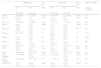
Changes associated with the PR program involving ACTSubjects as a wholeAfter ACT treatment, we observed significant improvements in most physiological measures both in the TPEP and PEP mask groups (Table 2). Almost all ventilatory parameters improved significantly in both groups, i.e., FVC and FVC%, FEV1 and FEV1% predicted, and peak expiratory flow and PEF% predicted. Most gas exchange parameters (SaO2, partial pressure arterial oxygen (PaO2), and ratio of partial pressure arterial oxygen and fraction of inspired oxygen (PaO2/FiO2)) were also greatly improved in both ACT groups, while partial pressure arterial carbon dioxide (PaCO2) remained unchanged.
Main results in the overall study group.
| TPEP group | Pre vs. post | PEP group | Pre vs. post | ΔTPEP vs. ΔPEP | |||
|---|---|---|---|---|---|---|---|
| Pre | Post | TPEP | Pre | Post | PEP | Intergroup comparison | |
| Mean (SD) | Mean (SD) | Mean (SD) | Mean (SD) | ||||
| FVC (L) | 2.06 (0.66) | 2.18 (0.66) | <0.001 | 2.03 (0.57) | 2.21 (0.70) | <0.001 | NS |
| FVC% | 69.44 (21.95) | 73.73 (21.93) | 0.015 | 71.15 (19.02) | 74.78 (23.72) | 0.015 | NS |
| FEV1 (L/s) | 1.16 (0.53) | 1.21 (0.54) | <0.001 | 1.16 (0.44) | 1.27 (0.52) | <0.001 | NS |
| FEV1% | 50.00 (23.46) | 52.22 (23.97) | <0.001 | 53.11 (21.59) | 58.41 (24.10) | <0.001 | NS |
| FEV1/FVC | 56.07 (13.39) | 55.02 (12.64) | NS | 57.79 (14.10) | 58.54 (15.33) | NS | NS |
| FEF25–75% | 22.31 (20.1) | 22.22 (17.31) | NS | 25.83 (20.09) | 30.80 (25.73) | NS | 0.05 |
| FEF50% | 21.84 (21.74) | 21.39 (18.94) | NS | 25.28 (21.51) | 29.85 (26.73) | NS | 0.046 |
| FEF75% | 22.48 (16.03) | 23.75 (17.28) | NS | 28.11 (23.06) | 31.95 (26.33) | NS | NS |
| PEF (L/s) | 3.46 (1.56) | 3.63 (1.42) | NS | 3.31 (1.22) | 3.88 (2.59) | NS | NS |
| PEF% | 52.39 (23.33) | 54.45 (20.25) | 0.006 | 51.26 (18.43) | 56.32 (20.59) | 0.006 | NS |
| SaO2% | 93.06 (3.92) | 94.31 (2.28) | <0.001 | 92.75 (3.41) | 94.45 (1.98) | <0.001 | NS |
| PaO2 (mmHg) | 64.7 (13.99) | 71.09 (13.76) | <0.001 | 65.24 (9.67) | 71.46 (11.85) | <0.001 | NS |
| PaCO2 (mmHg) | 43.96 (9.32) | 44.01 (7.90) | NS | 43.77 (9.72) | 43.67 (10.94) | NS | NS |
| PaO2/FiO2% | 274.21 (73.17) | 305.94 (83.78) | <0.001 | 289.33 (56.34) | 311.99 (70.42) | <0.001 | NS |
FVC (FVC%), forced vital capacity (% predicted); FEV1 (FEV1%), forced expiratory volume in one second (% predicted); FEF25–75(%), FEF50(%), FEF75(%), forced expiratory flows at 25–75%, 50% and 75% of FEV1 (% predicted respectively); PEF (PEF%), peak expiratory flow (% predicted); SaO2, arterial oxygen saturation; PaO2, partial pressure arterial oxygen; PaCO2, partial pressure arterial carbon dioxide; FiO2, fraction of inspired oxygen; PaO2/FiO2, ratio of partial pressure arterial oxygen and fraction of inspired oxygen.
Comparing the magnitude of the changes observed after the two types of treatment (ΔTPEP vs. ΔPEP), there were no significant differences apart from borderline FEF25–75% and FEF50% improvements in the PEP mask group (Table 2).
Subject subgroupsThe large number of subjects evaluated in the present study enabled us to perform analyses in subgroups to detect any specific effects of either of the two ACTs. We compared the changes observed after treatments in the two groups of ACT stratifying the subjects according to the categories reported in Table 1.
There were no differences between the effects of treatment with PEP mask or TPEP in terms of primary diagnosis (bronchiectasis vs. CB/COPD), smoking history, presence or absence of an acute exacerbation, or level of FEV1% predicted (> vs. <50%). Furthermore, treatments were equally effective in both treatment groups irrespective of their duration (< or >15 days), suggesting that 10 days of ACT (i.e., the shortest length of treatment allowed) are sufficient to yield a positive effect on clinical and physiological parameters regardless of the method used.
In subjects with emphysema, we observed some interesting differences between the two groups. The need for supplemental oxygen before and after differed significantly in TPEP subjects (reduced) compared to those treated with PEP mask (increased, p=0.034, Fig. 2A). In addition, the FiO2 at which the patients were breathing differed significantly between the PEP and TPEP groups (p=0.031, not shown). In this context, in the TPEP group, oxygen supplementation was reduced in the majority of subjects with emphysema, and one subject discontinued it completely. In the PEP group, many subjects increased the oxygen supplementation, and three of them who were not on oxygen at baseline started to use it. PaCO2 diminished only in the TPEP group, while it increased in subjects treated with PEP mask (p=0.02, not shown).
 Means and standard deviations of oxygen supplement in subjects with emphysema in the TPEP group (A) and PEP group (B). *p=0.034, comparison between intergroup changes. (Panel B) Means and standard deviations of oxygen supplement in subjects on oxygen therapy (OT) in the TPEP group (A) and the PEP group (B). *p=0.046, comparison between intergroup changes. O2 suppl, oxygen supplement is expressed in L/min, liters per minute; TPEPpre, baseline value in the TPEP group; TPEPpost, post-treatment value in the TPEP group; PEPpre, baseline value in the PEP group; PEPpost, baseline value in the PEP group.")
(Panel A) Means and standard deviations of oxygen supplement in subjects with emphysema in the TPEP group (A) and PEP group (B). *p=0.034, comparison between intergroup changes. (Panel B) Means and standard deviations of oxygen supplement in subjects on oxygen therapy (OT) in the TPEP group (A) and the PEP group (B). *p=0.046, comparison between intergroup changes. O2 suppl, oxygen supplement is expressed in L/min, liters per minute; TPEPpre, baseline value in the TPEP group; TPEPpost, post-treatment value in the TPEP group; PEPpre, baseline value in the PEP group; PEPpost, baseline value in the PEP group.
As shown in Table 1, the majority of our subjects were on OT and/or MV. Data referring only to all those on oxygen therapy showed that the amount of oxygen supply diminished significantly in the TPEP group compared to the PEP mask group (p=0.046, Fig. 2B). In addition, in the TPEP group, the FiO2 diminished in the majority of subjects on OT whereas it increased in the PEP group (p=0.038, not shown), and six subjects who were not on oxygen started to use it.
In subjects on MV, PEP mask was superior to TPEP in increasing FEF50 and FEF50%, TPEP treatment was associated with FEF50% reductions and PEP mask treatment with its increase (p=0.018, Fig. 3). A similar but not statistically significant trend was observed in the same subjects for the changes in FEF25–75% and FEF25–75%.
 and the PEP group (B). TPEPpre, baseline value in the TPEP group; TPEPpost, post-treatment value in the TPEP group; PEPpre, baseline value in the PEP group; PEPpost, baseline value in the PEP group. *p=0.018, comparison between intergroup changes.")
Means and standard deviations of FEF50%, percent of predicted forced expiratory flow at 50% of vital capacity, in subjects on mechanical ventilation in the TPEP group (A) and the PEP group (B). TPEPpre, baseline value in the TPEP group; TPEPpost, post-treatment value in the TPEP group; PEPpre, baseline value in the PEP group; PEPpost, baseline value in the PEP group. *p=0.018, comparison between intergroup changes.
Finally, in subjects without CRF (neither on OT nor MV) we observed statistically significant differences in PaO2 (p=0.0024) and PaO2/FiO2 (p=0.021) between groups. TPEP group showed a greater improvement of both PaO2 and PaO2/FiO2. Of note, in this subgroup of patients, baselines PaO2 values were rather low (<70mmHg).
DiscussionBy evaluating two groups of subjects with CH in the common clinical scenario, included in a standard PR program and treated with different ACTs, we found improvements in the PaO2/FiO2 ratio and in many important physiological parameters. Furthermore, our data show that TPEP performs as well as is the most commonly used PEP mask. Finally, subgroup analyses indicate that TPEP and PEP could perform differently in different conditions, suggesting that an accurate selection of ACTs, not exclusively tailored to individual preference or comfort, is warranted to obtain a better performance.
A previous report on the experimental use of TPEP showed improvements in symptoms, static and dynamic lung volumes, and a significant and progressive reduction in mucus production and in perceived bronchial encumbrance in chronic respiratory diseases, including COPD, asthma, and cystic fibrosis, and in the preparation of COPD patients for major abdominal surgery.20 Based on those preliminary findings suggesting an effect on the distribution of lung ventilation with significant improvements in ABGs, a multi-center randomized controlled trial was designed.13 The “UNIKO Project” showed that TPEP in combination with manually assisted ACT, namely ELTGOL21–23 (Expiration Lente Totale Glotte Ouverte en Décubitus Latéral) improves lung function and symptoms in subjects with CH and promotes a faster recovery from symptoms during treatment compared to ELTGOL alone. In that study, there were no significant differences in ABG values between the two groups.
Our analysis, performed on subjects admitted to the Pneumology Department of a rehabilitation center and conventionally treated with a comprehensive PR program including ACT techniques, confirms previous experimental results. In particular, it confirms that such an approach can produce significant improvements in respiratory function and ABGs in a mixed population of patients. However, our findings and previous observations18 are in contrast with the “UNIKO Project” report stating that the addition of TPEP did not produce any change in ABGs.
There are a number of differences between our study and the “UNIKO Project” that may explain the differences in the results observed. The study by Venturelli et al.13 had a number of exclusion criteria as it enrolled only subjects in stable conditions, excluding those with acute exacerbation or on MV. In fact, ABG values at the beginning of their treatments were normal, so finding significant changes after treatment would have been very unlikely. Despite this, their TPEP group still showed a better trend in a shorter period of treatment.
In the present study, we evaluated a larger number of subjects in a “real-life” clinical scenario. They were consecutive subjects with CB/COPD or bronchiectasis as primary diagnosis and history of CH who were enrolled in a PR program including ACT. Thus, we included a significant percentage of subjects with acute exacerbations and those on OT and/or MV (39, 47, and 19%, respectively). In addition to our comparison, we differentiated the two types of ACT (TPEP vs. PEP), while Venturelli et al.13 added TPEP to conventional treatment.
Our results show that on average both groups had equally improved physiological parameters such as lung volumes, flows, and ABG values. Both PEP-mask and TPEP are classified as PEP methods aimed to favor the mobilization of secretions in the lung by “stenting” the airways. The mechanism and effects of PEP are widely described in the literature. PEP increases intrathoracic pressure and FRC, with improvement in collateral ventilation.24 The application of a 15cmH2O PEP temporarily increases VT, reducing respiratory frequency,25 and both inspiratory and expiratory muscle activity.26 In addition, a temporary increase in FRC, related with increasing PEP has been shown.27 This leads to changes in breathing pattern, with decreased expiratory flows, increased expiratory time, and a smaller exhaled volume.28 PEP pressures higher than 10cmH2O are required to open the airways.12
With this in mind, it seems difficult to understand how a pressure as little as 1cmH2O, such as that generated by TPEP when measured at the mouth, could be as effective as the PEP mask. Other factors probably play a role. In fact, in the TPEP device, there is a pressure sensor that detects exhalation, activating a compressor that blows air against the expiratory flow. The resistance delivery time depends on the ability of the subject to sustain a certain flow as long as possible. A forced exhalation will generate a peak of expiratory flow causing a sudden drop in pressure (that leads to a collapse of the peripheral airways). The best performance is obtained when the expiratory flow is a little higher than during quiet breathing, almost reaching residual volume and achieving the same goal as that of other classical ACTs such as autogenic drainage and ELTGOL.22,29 Another difference between the two ACTs is that, with the PEP mask, the flow is variable and effort-dependent,24,30,31 while the TPEP device gives feedback and guides the subject to adapt inspiration and expiration to reach the goal. Furthermore, while blowing into the mouthpiece, an oscillation is clearly perceived while resistance is active, due to the reciprocating compressor. High-frequency oscillations are thought to have a mucolytic effect on bronchial secretions,32 and this feature could play some role in explaining the mechanism of this technique.
Operational differences between PEP mask and TPEP may also account for the differences found between the effects obtained by the two ACT techniques in our study. Subgroup analysis gave some preliminary indications. Subjects with emphysema showed a reduced need for oxygen (O2) supply, FiO2, and PaCO2. The above data, combined with the general reduction in the need for O2 in OT subjects and with the increase in PaO2 in those without CRF, seems to indicate that TPEP may facilitate gas exchange. During TPEP, very low pressures are applied to the airways, making unlikely any direct recruitment effect or changes in surfactant activities and/or production as seen with PEP systems.30
We speculate that breathing pattern and the effective mucus clearance achieved by the increase in gas–liquid interaction33 during sustained expiratory flow could explain gas-exchange improvements. It would also be interesting to investigate if any post-treatment change is present in respiratory pattern and mechanics or in airway resistance. In fact, TPEP forces patients to breathe in a regular way, with open glottis, for the entire treatment time, and this “training” could have contributed to improve the ability to control breathing in subjects with emphysema.26,34
We observed a statistically significant difference between groups when evaluating subjects on MV only for the forced expiratory flows (FEF50%). The interpretation of this finding is difficult particularly because volumes or gas exchange parameters did not differ between subjects on MV. We might speculate that MV, providing positive pressure inside the airways, can overcome the TPEP effects on FEF. On the contrary, the administration of a higher PEP with FET maneuvers could have enhanced mid-expiratory flows. Clearly, further analyses are needed to confirm this hypothesis.
Comfort is another important factor we take into account when allocating patients to different treatments. Notably, many of the subjects treated with TPEP were already PEP-mask users at home and found the new device more comfortable and simple to use, since less effort is required compared to the PEP mask with the same subjective perception of efficacy. This feedback was important, prompting us to continue investigations and, as underlined by many authors,10,11,35–38 it might improve adherence to the treatment.
The major limitation of the study is the retrospective design and other sources of bias such as the consequent lack of randomization and intention-to-treat analysis. Thus, caution should be applied when interpreting the results of this study. Moreover, future randomized controlled trials are warranted to confirm the results of this study.
Other limitations include the lack of data on the volume and characteristics of sputum, symptoms and health-related quality of life, not systematically available in the clinical documentation for all patients included. Furthermore, allocation to one or other treatment group may have been affected by the patient's perception and compliance during the first assessment. TPEP requires less effort than PEP to be activated: this might explain in part why a greater proportion of subjects with an acute exacerbation were in the TPEP group, though no significant differences were found between the effects of treatment in relation to the presence of an acute condition.
Nevertheless, no other differences in baseline parameters were found between groups. Furthermore, the PEP mask is a well-established technique that physiotherapists and patients are familiar with, while at that time, TPEP had just been introduced into clinical practice, a fact that probably was the cause of the smaller number of subjects treated with this newer technique.
ConclusionsIn conclusion, our data showed that, in most subjects with CH in current clinical practice, TPEP yields similar results to the PEP mask. Both groups significantly and similarly improved physiological parameters, but subgroup analysis suggests that TPEP could provide some advantages to subjects with emphysema or those on OT, while PEP would be more advantageous in patients on MV. TPEP added to a standard PR program applied in subjects with CH appears to be a safe and well-accepted ACT and a valid alternative to PEP. We need further prospective, high quality randomized controlled trials to confirm preliminary results indicating that different subgroups/phenotypes can benefit more from one or the other type of treatment.
Conflicts of interestThe authors declare no conflict of interest.


