


Femoroacetabular impingement syndrome (FAIS) is a hip joint motion-related clinical disorder with a triad of symptoms, clinical signs, and imaging findings. However, scientific evidence is still unclear regarding the best treatment for FAIS.
ObjectivesTo assess the value of a physical therapy evaluation in predicting the progression of functional status over the subsequent years in patients with FAIS who are candidates for hip arthroscopy surgery.
MethodsIn this case-series study, patients with FAIS, candidates for hip arthroscopy surgery, underwent a standard physical therapy evaluation. Baseline data were collected between 2013 and 2019. In 2020/2021, the patients’ functional status was assessed through the International Hip Outcome Tool (iHOT-33). Functional status progression was calculated as the difference between the follow-up and baseline iHOT-33 scores. A multivariate forward stepwise regression analysis was conducted to explore the relationship between baseline characteristics and the functional status progression.
ResultsFrom 353 patients who completed the baseline assessment, 145 completed the iHOT-33 follow-up. The mean (±SD) follow-up time was 58.7 (27.2) months (minimum 12 and maximum 103 months). The iHOT-33 scores increased 20.7 (21.8) points on average, ranging from -39.8 to 76.9 points. Among the 15 potential predictive factors assessed in this study, only baseline iHOT-33 score (β -0.44; -0.061, -0.27), femoral version (β 9.03; 1.36, 16.71), and body mass index (β -0.99; -1.98, -0.01) had the ability to predict the functional status progression.
ConclusionPatients with a lower baseline iHOT-33 score, lower body mass index, and normal femoral version were more likely to increase their functional status after a minimum of one year of follow-up.
Femoroacetabular impingement syndrome (FAIS) is a hip joint motion-related clinical disorder with a triad of symptoms, clinical signs, and imaging findings.1 The impingement between bone structures causes cartilage damage, pain, and predisposes the hip to early osteoarthritis.2 Scientific evidence is still unclear regarding the best treatment for FAIS.3,4,5 The clinical decision to undergo conservative or surgical treatment usually depends on subjective aspects related to pain intensity and the impact on the patient's quality of life. Therefore, patient-reported outcome measures (PROMs) play a crucial role in evaluating the impact of FAIS progression from the patient's perspective and have been extensively utilized to aid in clinical decision-making.6
The International Hip Outcome Tool (iHOT-33)7 was recommended as one of the most appropriate PROMs to be used in young and middle-aged active adults with hip-related pain.6 High-quality randomized controlled trials involving patients with FAIS have included the iHOT-33 score as the primary outcome measure.3,4 In one of the most extensive trials, Griffin et al.3 observed that arthroscopy and physical therapy approaches led to notable improvements in patients' iHOT-33 scores, with an average increase of approximately 20 and 14 points after 12 months, respectively. While most patients have a positive prognosis regardless of the treatment option, there still exists uncertainty about identifying which patients are more likely to achieve a favorable outcome.
There is some confusion about the definitions of the terms “prognostic” and “predictive” in the literature. A prognostic factor is a characteristic that provides information on the likely outcome of the disease in an untreated individual.8 In contrast, a predictive factor is a characteristic that provides information on the likely benefit from treatment.8 In case of FAIS, evidence suggests that young men, with body mass index (BMI) <24.5 kg/m2, a Tönnis grade of 0, and pain relief from preoperative intra-articular hip injections are more likely to achieve positive outcomes after hip arthroscopic surgery.9 Conversely, as far as we know, there is a lack of investigations into predictive factors for patients undergoing conservative treatment. Moreover, predictive value studies have failed to consider variables such as muscle strength and range of motion (ROM). Given that patients with FAIS typically exhibit hip muscle weakness and reduced hip ROM compared to healthy individuals,10,11 it is plausible to speculate that muscle strength and/or joint mobility may play a role in determining the course of their functional status. Likewise, considering that femoral version (FV) abnormalities are known to influence the development of hip pain and can also negatively impact both the strength and ROM in patients with FAIS,12,13 the predictive value of FV warrants further investigation.
Determining the factors identified during a clinical evaluation that can predict a higher likelihood of improvement or deterioration in the functional status of patients with FAIS undergoing surgery or conservative treatment is of paramount importance. Hence, the primary objective of this investigation was to assess the value of a physical therapy evaluation in predicting the progression of functional status over the subsequent years in patients with FAIS who are candidates for hip arthroscopy surgery.
MethodsStudy designThis is a case series. First, information was retrospectively obtained from records of patients with FAIS evaluated between 2013 and 2019 at the Physique Physical Therapy Clinic (Porto Alegre, RS, Brazil), which is considered a reference center for the rehabilitation of hip-related disorders. In the prospective phase of this study, conducted between November 2020 and July 2021, eligible patients who were a minimum of one year post initial evaluation were invited to complete the iHOT-33.14 Fifteen variables were assessed as potential predictive factors on the functional status progression of patients with FAIS, represented by the change in iHOT-33 scores from baseline to follow-up evaluations. The study was approved by the Ethics Committee from the Universidade Federal de Ciências da Saúde de Porto Alegre (1.871.372). All participants signed an informed consent form agreeing with future use of data for research.
ParticipantsAll participants were evaluated by an orthopedic surgeon and diagnosed with FAIS based on the following criteria: presence of motion-related or position-related pain symptoms at the hip; pain and limited ROM with the Flexion Adduction Internal Rotation (FADDIR) hip impingement test; and morphological abnormalities determined on imaging.1 An anteroposterior radiograph of the pelvis and a lateral neck view were performed to determine hip morphological abnormalities. All patients were candidates for hip arthroscopy surgery.
The study excluded patients: younger than 17 years old or with a history of prior hip surgery; with diabetes mellitus; with neuromuscular, neurological, or rheumatological diseases; as well as those with missing data from the initial physical therapy evaluation. Contacted patients were included if they completed the iHOT-33 questionnaire at follow-up or excluded if they did not. Baseline values from both groups were compared to determine the groups’ similarity at baseline.
We estimated that at least 112 patients would be needed to evaluate the linear correlation between the iHOT-33 delta and a selected group of potential predictive factors. We used a two-tailed Type I error level of 0.05 and a statistical power of 90%, assuming a minimum relevant correlation of 0.3. To account for the number of variables tested and adjustments in the multivariable regression model, the sample size was increased to 140 patients.
Primary outcomeThe primary outcome was the iHOT-33 delta score, calculated as the difference between the follow-up and baseline scores. Patients completed the iHOT-33 tool in the clinic for the initial physical therapy evaluation and used the REDCap online platform for the follow-up evaluation.15,16 iHOT-33 presents adequate psychometric properties according to a consensus statement from the IHiPRN meeting.6 iHOT-33 has shown acceptable construct and content validity, good reliability (ICC = 0.78–0.88), and minimal detectable change values ranging from 2.3 to 3.7 points in patients who were not seeking surgery.17 In addition, the minimal clinically important difference (MCID) for iHOT-33 is 10 points.18
Physical therapy evaluationAll evaluations were conducted by three physical therapists from the physical therapy clinic where the study was conducted. They had at least 10 years of experience in the management of patients with FAIS and received specific training to conduct the evaluation routinely employed in the clinic for this population, including the assessments described next.
ROM assessment: Hip joint ROM was assessed using a digital goniometer (Medigauge, Columbia, USA) for active flexion (FLX), internal rotation (IR), and external rotation (ER).19 For the measurements of hip FLX and ER, patients were positioned in supine with the contralateral leg in neutral and fully supported on the table. For IR, patients were in supine with both hips and knees flexed to 90°, and the movement was performed bilaterally and simultaneously.19
Femoral version assessment: FV was clinically assessed using the rotation index, a ROM-based measurement calculated by subtracting hip IR from hip ER ROM values.13 The rotation index has been shown to be an independent FV predictor in patients with hip pain: the probability of hip being anteverted or retroverted is greater than 70% if the rotation index is less than 11° or higher than 40°, respectively.13 Therefore, a normal FV was considered when the rotation index was between 11° and 40°, and FV was considered a dichotomous variable (i.e., normal or abnormal) in this study's prediction model.
Muscle strength assessment: Maximal isometric hip strength was assessed with hand-held dynamometry (MICROFET – Hoogan Scientific, Salt Lake City, USA). Hip ABD and ADD was assessed in side lying, while ER and IR were assessed in a seated position.19 In all tests, the dynamometer was placed distally on the leg, close to the ankle joint. For each muscle group, patients completed 2 valid maximal voluntary isometric contraction (MVIC) trials. If the difference between the 2 MVICs was greater than 10%, a third trial was performed. Participants received standardized verbal encouragement to produce maximal effort. A 120-s rest time was used between trials. The highest valid force measurement was converted to a hip joint torque value normalized to body mass (Nm/kg).19
Potential predictive factorsOverall, 15 variables were assessed as potential predictive factors. Thirteen factors were assessed at the initial physical therapy evaluation: age; sex; BMI; sedentary profile (determined by the question: do you do any regular exercise type or sports practice?); hip ROM for FLX, ER, and IR; FV; hip strength for ABD, ADD, IR, and ER; and baseline iHOT-33 score. The other two factors were obtained during follow-up evaluation: treatment option and time of follow-up. Although all patients were candidates for surgery, some patients decided to not undergo surgery; thus, we included treatment option as a confounder variable in the model. The time of follow-up was considered the time between the initial physical therapy evaluation and the follow-up evaluation.
Treatment optionsHip arthroscopies were performed at a local hospital that follows high quality standards using a technique previously described in the literature.20 The description of the surgery using a conventional optical 30° arthroscope was obtained from the surgical records dictated by the surgeons. As the first step, the capsular fatty tissue was cleaned, and a longitudinal capsulotomy along the femoral neck was performed using a 3.5 mm, 90° hook electrode. The proximal landmark was the indirect head of the rectus femoris muscle, where a T-shaped capsule extension was added whenever needed. A traction suture was passed through the capsular flaps to facilitate the exposure, preserving the stabilizing function of the iliofemoral ligament. Hip arthroscopy was then conducted in a routine manner by performing a femoral osteochondroplasty to remove the CAM morphology, which was performed on 100% of the patients. Labrum stability was then assessed with a probe. Confirmation of the required resection was done with fluoroscopy and dynamic visual assessment by performing the impingement test. When a pincer morphology was addressed, the labrum was detached to resect the acetabular rim, which was usually at the supra-equatorial portion of the acetabulum. The labrum was then re-attached using 2 or 3 suture anchors with the simple loop stitch technique.
All patients received instructions regarding post-operative care and the physical therapy protocol used at the clinic (Appendix 1). Also, a week-based exercise protocol was provided to the patients. This program was composed of specific exercises to gain mobility, improve muscle activation, and progressively improve muscle strength. For patients who did not have a confirmed surgery date, instructions were given regarding exercises and positions that should be avoided until the surgery, such as avoiding hip flexion greater than 90°, not performing stretches that forced the hip joint in extremes of movement, and avoiding high demand sports for the hip such as playing soccer and tennis.
Statistical analysisAll baseline characteristic values are presented as mean and standard deviation (SD). Continuous variables were compared with t-tests and categorical variables with chi-square test when needed. Potential predictive factors were entered into a multivariable forward stepwise regression analysis. Linear regression was used with the iHOT-33 delta score as the primary outcome. Associations within the multivariable model were regarded as significant at p < 0.05. The strength of the predictive value of identified factors was determined using unstandardized regression coefficient.
ResultsFrom the 523 patients with FAIS identified in the clinic's database, 353 were eligible for this study (Figure 1). From the eligible participants, 145 completed the follow-up phase. Patients included (those who completed the iHOT-33 at follow-up) and excluded (those who did not complete the iHOT-33 at follow-up) had similar baseline characteristics (Table 1). The mean follow-up time was 58.7 (27.2) months (minimum 12 and maximum 103 months).

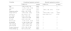
Baseline characteristics of the participants who answered (included) and did not answer (excluded) at follow-up.
Values are presented as n (%) or mean (SD). ABD, abduction; ADD, adduction; BMI, body mass index; CI, confidence interval; ER, external rotation; FLX, flexion; IR, internal rotation; ROM, range of motion; SD, standard deviation. *=chi-square p-value.
The average (SD) increase in the iHOT-33 score between evaluations was 20.7 (21.8) points (minimum −39.8; maximum 76.9 points). Ninety-nine (68.3%) patients enrolled in this study achieved an improvement in the iHOT-33 score of ≥10 points (i.e., the MCID for the iHOT-33).18 One hundred and four patients (71.7% of participants) underwent hip arthroscopy surgery, while forty-one (29.3%) patients decided to manage their condition without surgery. The average (SD) increase in iHOT-33 delta score found for patients undergoing hip arthroscopy and conservative treatment were 21.4 (23.9) points and 19.1 (15.8) points, respectively.
The univariate and multivariate forward stepwise regression analysis is presented in Table 2, showing that only baseline iHOT-33 score (β −0.44; p < 0.001), BMI (β −0.99; p = 0.047), and FV (β 9.03; p = 0.021) had the ability to predict iHOT-33 delta score in the multivariate model. A normal FV increased the iHOT-33 delta score by 9 points, whereas greater baseline BMI was associated with lower iHOT-33 delta scores. This model explained 20% of iHOT-33 delta score variance.
Univariate and multivariate forward stepwise regression analysis.
ABD, abduction; ADD, adduction; BMI, body mass index; CI, confidence interval; ER, external rotation; FLX, flexion; FV, femoral version; iHOT-33, International Hip Outcome Tool; IR, internal rotation; ROM, range of motion.
The present study showed that baseline iHOT-33 score, BMI, and FV can explain 20% of iHOT-33 delta score variance. Patients with a lower initial iHOT-33 score, a lower BMI, and a normal FV were more likely to increase their functional status at follow-up. Other variables evaluated during the initial physical therapy assessment, such as hip muscle strength and ROM, as well as the subsequent choice of surgery or conservative treatment, showed no significant association with the iHOT-33 delta score.
The analysis of the follow-up data revealed that almost 30% of the patients opted not to proceed with the surgery for which they were referred at the time of the initial physical therapy evaluation. As a result, we incorporated the treatment option as an intervening variable in the study, recognizing it as a potential predictive factor during the statistical analysis. Interestingly, the treatment option did not appear to significantly influence the functional improvement of patients. This finding suggests that the association analysis conducted for other potential predictive factors in our study remains unaffected by the patient's treatment option. In addition, the increase of delta iHOT-33 scores in conservative [19.1 (15.8) points] and surgical [21.4 (23.9) points] treatments is consistent with previous evidence that both conservative and surgical treatments lead to improvement in functional status.21
According to a systematic review by Sogbein et al.,9 young age, male sex, and lower BMI were found to predict a positive outcome after hip arthroscopy for FAIS. In our study, which included men and women from 17 to 65 years old, the patients' sex and age were not predictors of change in functional status. Regarding age, we speculate that the limited number of patients aged over 50 years (specifically, 14 participants, which is less than 10% of the sample) presents a limitation in assessing the influence of age on the iHOT-33 delta score.
As found by Sogbein et al.,9 BMI has been described as a predictive factor for patients with FAIS undergoing hip surgery. A BMI less than 24.5 kg/m2 was associated with improvement exceeding the MCID on the hip outcome score (HOS).22 Considering iHOT-33 delta score as an outcome, our results agree with these results, showing that greater BMI is associated with lower change in iHOT-33 delta score, regardless of the choice for surgery or conservative treatment. This result agrees with a recent systematic review,23 which showed that obesity is associated with worse pain and complications on clinical outcomes post hip and knee arthroplasty in patients with osteoarthritis. Because BMI is a modifiable factor, patients should be informed about the possibility of obtaining higher increments in iHOT-33 delta scores through weight loss strategies.
Considering that physical inactivity has been associated with chronic musculoskeletal complaints,24 it is reasonable to expect that a sedentary lifestyle may impact the prognosis of patients with FAIS. Fifty patients (34.5%) in our study reported a sedentary lifestyle at the initial evaluation, and our results supported that being sedentary was not a factor related with a lower iHOT-33 delta score. However, we did not assess if these patients were sedentary due to pain, or simply had a sedentary lifestyle, and if they continued to be sedentary during the follow-up period. These factors could have impacted our results. Based on the limitations of our analysis regarding sedentary lifestyle, we can only state that being sedentary at the time of initial physical therapy evaluation was not found to be a predictor of functional status in patients with FAIS.
Our analysis did not reveal any correlation between modifiable impairments, such as strength and ROM, and the iHOT-33 delta score. As far as we know, this study is the first to look at hip ROM as a potential predictive factor of functional status in patients with FAIS. Conversely, Beck et al.25 conducted the only study to investigate the predictive value of hip muscle strength in 74 patients with FAIS undergoing hip arthroscopy. According to their findings, maximum isometric strength for hip extension was a significant predictor of achieving meaningful outcomes at 6 months post-surgery.25 However, no predictive value was observed for hip rotators and abductors strength in their study.25 Our study findings align with those of Beck et al.,25 indicating that hip rotators and abductors strength did not serve as predictors of the change in functional status in patients with FAIS.
The modifiable nature of variables such as strength and ROM might explain their poor predictive value in a long-term follow-up study. Over the years, patients may improve their muscle strength and joint ROM due to pain reduction, increased exercise level, and improved general health status, or conversely show further decline due to worsening of symptoms and a sedentary lifestyle. As we did not measure hip strength and ROM during the follow-up period, future investigations should assess the predictive value of hip strength and ROM and changes from baseline to the follow-up periods. In addition, given the large variability in follow-up period from baseline evaluation, it is possible that numerous other factors not accounted for in our study might be associated with our outcomes (as evidenced by only 20% of the variance in iHOT-33 delta scores being explained by our final model).
FV has been recognized as a potentially important factor in the development of hip pain.26 Because 52% of the hips scheduled for hip preservation surgery have abnormal FVs,26 and patients with abnormal version had less improvement after hip arthroscopy than those with a normal version angle,12 FV may also be an important factor related to health prognosis in patients with FAIS. According to our results, 111 (77%) patients presented a rotation index indicative of a normal FV. Additionally, patients with normal FV had a 9-point greater increase in the iHOT-33 delta score. Therefore, in addition to being a feasible tool to predict FV in the clinical setting,13 rotation index is suggested to be a valuable measure for predicting changes in functional status of patients with FAIS following conservative and surgical treatments.
This study has some limitations. First, the time between evaluations ranged from 1 to 8 years. This variability may have determined different iHOT-33 scores in the follow-up evaluation, although self-reported pain and function seems to reach a plateau between 12 and 24 months after surgery.27 Second, although all patients were candidates for surgical treatment, almost 30% decided for conservative approaches. The low number of patients (n = 41) opting for conservative treatment did not allow us to evaluate predictive factors in each treatment group. We understand that pooling these interventions together may confound associations between variables, although we tried to minimize this by including treatment option as a variable in the model. Finally, the high number of patients who did not complete the follow-up iHOT-33 may result in selection bias, although there was no difference in baseline characteristics between those who completed the iHOT-33 and those who did not.
ConclusionPatients with FAIS with lower baseline iHOT-33 score, lower BMI, and normal FV were more likely to increase their functional status after a minimum of one year of follow-up regardless of treatment option (surgery or conservative care). Physical therapists can take these predictive factors into consideration and utilize this information to align the expectations of patients with FAIS with their most probable outcome. This informed approach can help optimize treatment plans and provide patients with a better understanding of their potential progress.
During the postoperative recovery, a rehabilitation program will be performed, which has been previously discussed with your surgeon. Below are some details of the program.
- ✓
Immediate start, after hospital discharge.
- ✓
Frequency: physical therapy at the clinic (2 to 3 times a week) and daily exercises at home for 5 weeks.
- ✓
Use of continuous passive movement device for 1 h in the first two weeks.
- ✓
The protocol follows an exercise routine aimed at: improving muscle activation, improving joint mobility, progressive muscle reinforcement, and improving neuromuscular and proprioceptive control.
- ✓
After this 5-week physical therapy protocol, you will be released for exercise at the gym with supervision.
- ✓
The exercises may cause some discomfort in the operated hip, but it should not cause excessive pain.
- ✓
Apply ice to the operated hip for 20 min, 5 times a day, during the first 5 postoperative days.
Daily life activities:
- ✓
Weight bearing as tolerated with crutches for 15 days.
- ✓
Vertical stationary bike: 15 days after surgery.
- ✓
Driving: 10 days to 2 weeks after surgery.
- ✓
Stairs/curb: in an adapted way, it can be performed immediately after hospital discharge (climb the step first with the non-operated limb and descend the step first with the operated limb).
- ✓
If you have stairs at home, organize yourself to avoid excesses.
- ✓
Avoid sitting for long periods (maximum = 30 min). Alternating positions (sitting, standing, short walks, and lying down) is healthy for your recovery.
- ✓
Sleeping position: on your back, on your stomach, or on your side (on your non-operated hip with a pillow between your legs).
Physical activity and sports:
- ✓
Exercises at the gym: 5 weeks after surgery
- ✓
Treadmill running: approximately 3 months after surgery
- ✓
Street running: approximately 4 months after surgery
- ✓
Elliptical trainer: 2 months after surgery.
- ✓
Swimming: 1st month after surgery (crawl and backstroke only). You should not use breaststroke and dolphin/butterfly.
- ✓
Return to sport: gradual return starting the 5th to 6th month after surgery.


