

Muscle mechanical properties (MMPs) are relevant in the pathophysiology of lumbopelvic disorders. However, they have not been described in the pelvic floor muscles (PFM) and lumbar paravertebral muscles (LPM) of women with urge urinary incontinence (UUI).
ObjectiveTo identify differences between MMPs of PFM and LPM in patients with UUI and healthy controls. Secondarily also aimed to observe the relationship between sociodemographic and clinical variables with the PFM and LPM MMPs.
MethodsThe participants of this case-control study comprised 34 women with UUI (UUI group) and 34 continent women (control group). Sociodemographic variables were obtained together with data on the clinical status of the pelvic floor. The MMPs, i.e., frequency (tone), stiffness, decrement (inverse of elasticity), and viscoelastic properties (VP), such as relaxation time and creep, of PFM and LPM were assessed with a hand-held tonometer. Between-group differences and intra-group correlations were identified.
ResultsThe UUI group presented higher frequency and stiffness, as well as lower relaxation time in PFM, whereas the LPM had lower tone and stiffness, and higher VP, compared to the control group (p < 0.05). The UUI group showed a pattern of moderate correlations (|0.403|
ConclusionThe presence of UUI may influence MMPs at PFM and LPM levels, increasing the tone and stiffness of PFM, whereas these properties are reduced in LPM. These findings emphasize the clinical interest of the lumbopelvic determination of MMPs, obtained through externally applied hand-held instruments, in the pathophysiology of UUI.
The pelvic floor (PF) is essential for numerous functions such as pelvic girdle stability, continence, urination, defecation, sexual function, and childbirth.1 Deficits in the function of the pelvic floor muscles (PFM) or damage to the PF structures can compromise their action and result in urinary incontinence (UI). Due to its economic impact on health services, UI is considered a social problem,2 and this condition is aggravated by its high prevalence rates, mainly in women, with approximately 51.1 % reporting experiencing UI.3 In addition, the incidence of urge urinary incontinence (UUI) or mixed urinary incontinence (MUI) appears to increase with age.4 Although less frequent, women with UUI report a sudden urge to urinate ("urgency") followed by immediate leakage of urine, which results in a greater impairment of quality of life compared to patients with stress urinary incontinence.5 Despite the significant social and psychological repercussions of UUI, its etiology is not well understood, and it remains difficult to treat.5,6
The underlying cause of UUI appears to be multifactorial and includes abnormalities in bladder receptors, peripheral and central innervation, alterations in the PFM, and behavioral factors.7,8 Among these factors, alterations in the activity of the PFM, such as changes affecting its motor control, are considered some of the most relevant disorders requiring treatment to preserve continence.9,10 Along these lines, previous studies have observed possible relationships between UUI and impaired PFM strength and function, as well as decreased perception and pressure of muscle contractions.11,12 Moreover, different authors have assessed the importance of analyzing the tissue mechanical properties of the PF in pelvic floor disorders (PFD).13–15 Nonetheless, information regarding muscle mechanical properties (MMPs) in women with UUI is scarce and is sometimes obtained with non-objective methods such as digital palpation, which may not specifically identify MMPs.16
In the past few years, tonometry has helped to assess MMPs in different regions and clinical states,13,17 and has shown relevant relationships between MMPs and other clinical features.14,18 Likewise, the analysis of the MMPs of the lumbar musculature has also been of great interest in the pathophysiology of different pathologies.19 Tonometry consists of recording the damped natural oscillation of soft biological tissue, after exerting mechanical pulses over a compression force, in the form of an acceleration signal and the subsequent simultaneous computation of different parameters, such as state of tension or stiffness.20 Manual tonometry, one of the most used methods to assess MMPs, has been shown to be a valid,21 reliable,22 noninvasive, and safe option of assessing MMPs, in contractile23 and non-contractile tissues,24 especially useful in clinical settings.
Previous studies have described relationships between lumbopelvic stability, spinal mobility, and low back pain, with PF dysfunctions,25,26 including UUI.25,27 Nonetheless, despite the relevance of the study of the lumbar spine in PF dysfunctions, because of its possible regional interdependence,28 there is no information on the MMPs of the lumbar paravertebral muscles (LPM) in patients with UUI. Therefore, there is a need for greater knowledge of the lumbar MMPs to determine their involvement in the pathophysiology of UUI and to improve the management of associated symptoms.
Therefore, the objective of this study was to identify differences between MMPs of PFM and LPM in patients with UUI and healthy controls. Secondarily the study also aimed to observe the relationship between sociodemographic and clinical variables with the PFM and LPM MMPs.
MethodsStudy designA case-control study was conducted following the recommendations of the STROBE Declaration. This study complied with the Declaration of Helsinki and Ethical approval was obtained from the Research Ethics Committee of Córdoba, Spain (code 4074, 2018). In addition, all participants signed the informed consent form prior to their participation in this study.
SubjectsA total of 68 women between 18 and 65 years of age participated and were divided into two groups according to a prior UUI diagnosis by a physician and the result obtained on the Three Incontinence Questions (3IQ).29,30 Thus, 34 women with a previous diagnosis of UUI were assigned to the UUI group, and 34 controls without UUI were included in the control group. Both groups were matched for age (±5 years) and BMI (±3 kg/m2). Recruitment was performed through social networks and flyers posted on the university campus. Exclusion criteria were: BMI greater than 40 kg/m2; known anatomical alterations in the PF or surgery in the lumbopelvic area; receiving medical treatment or seeking medical treatment for UUI that interfered with the status of the PF tissue characteristics; presence of scoliosis; low back pain in the last 6 months; any systemic disease that interfered with the anatomy and physiology of the PF.
To identify a minimum detectable change of 0.86 Hz, with a pooled standard deviation of 1.23 Hz for Frequency,22 an error Type I of 0.05, and power of 0.80, 34 women per group were necessary (G*power 3.1.9.2, t-test for Difference between two independent groups procedure).
ProceduresAfter signing the written informed consent, sociodemographic and clinical data were collected from the participants using the validated Spanish version of the Pelvic Floor Distress Inventory (PFDI-20) and the Pelvic Floor Impact Questionnaire (PFIQ-7), that have shown intraclass correlation coefficients of 0.644 and 0.786 for test-retest reliability, respectively, and Cronbach's alpha coefficients higher than 0.8 for internal consistency.31,32 For both questionnaires, the total score ranged from 0 to 300, where higher scores mean high distress and impact, respectively. Finally, the level of physical activity for each woman was categorized as low, moderate, or high, according to the Global Physical Activity Questionnaire (GPAQ),33 that has shown good-to-very good test–retest reliability (Pearson r coefficient = 0.58–0.89).34
Assessment of the MMPs of the PFM and, subsequently, of the LPM was performed with the MyotonPRO device (Myoton AS, Tallinn, Estonia), following standardized measurement protocols and with an empty bladder.22 For the assessment of the MMPs of the PFM, the volunteers were asked to lie supine on the treatment table, with their knees bent and the soles of their feet on the table. The participants were instructed by verbal command to perform a maximal voluntary PFM contraction prior to measurement, facilitating subsequent muscle relaxation. The measurement site was located on both sides of the central core of the perineum, located by visual observation and palpation at the largest area of muscle mass during contraction.15,22 Subsequently, the participants were asked to lie prone on the table for the assessment of the MMP of LPM, the evaluator located the spinous process of the L4 vertebra and measured both sides starting with the right side.19 In both regions, recording was performed during five seconds of apnea after exhalation to reduce the abdominal influence on the test.21
The MyotonPro provided data for five parameters: frequency (Hz), representing the tonus or state of active tension, stiffness (N/m), logarithmic decrement (the inverse of elasticity), and the viscoelastic properties (VP), relaxation time (ms), and creep (representation of the Deborah number).15
A visual analogue scale (VAS) was used at the end of the assessments to identify the presence of pain during the protocol.22 Testing took less than 30 min.
Statistical analysisCategorical variables were presented as frequencies and percentages, and continuous data as means and standard deviations with a 95 % confidence interval (95 % CI). The Kolmogorov–Smirnov test showed a normal distribution (all variables: p > 0.05, with the exception of PFDI-20 and PFIQ-7, that were described with median and interquartile range). For both the PFM and the LPM, the mean values combining both sides was used for the analyses, because preliminary analysis indicated that there were no differences between sides (paired Student-t-test, p > 0.05).
To identify between-groups differences in MMP and sociodemographic variables, unpaired Student-t and Mann-Whitney-U tests were conducted, depending on normal distribution.
To determine intra-group associations between the MMPs and sociodemographic and clinical data, Pearson's r and Spearman's rho coefficients were calculated. Correlations were considered to be negligible (0.0 to 0.19), fair (0.20 to 0.39), moderate (0.40 to 0.69), strong (0.70 to 0.89), or almost perfect (0.90 to 1.00).35
The level of significance was set at 0.05. The IBM-SPSS® software, version 25 (SPSS Inc., Chicago, IL, USA), was used for the analyses.
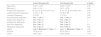
ResultsThirty-four women were included in each group. For the sociodemographic and clinical data, only the results of the questionnaires showed statistically significant differences between groups (p < 0.05). Both groups included young adult women with similar variability in age, and presented mean normal BMI values (20–25 kg/m2). Up to 56 % of women with UUI and 62 % of controls experienced at least one pregnancy; 50 % and 62 %, respectively, had a previous vaginal delivery. The number of women undergoing menopause was higher in the UUI group compared to the control group (8 and 6, respectively). No woman expressed pain or discomfort due to the examination (VAS=0). Additional data are shown in Table 1.
Sociodemographic and clinical data of the control and urge urinary incontinence (UUI) groups.
| Control Group(n=34) | UUI Group(n=34) | p-Value | |
|---|---|---|---|
| Age (years) | 37.97 ± 12.68 | 34.65 ± 13.55 | 0.30 |
| BMI (kg/m2) | 22.61 ± 3.16 | 24.71 ± 5.79 | 0.05 |
| Pregnancies (frequency) | 0: 13; 1: 6; 2: 10; 3: 54: 0; 5: 0 | 0: 15; 1: 2; 2: 10; 3: 54: 0; 5: 2 | 0.38 |
| Vaginal deliveries (frequency) | 1: 9; 2: 11; 3:1 | 1:2 2:10; 3:5 | 0.05 |
| Cesarean (frequency) | 1: 3; 2: 2 | 1: 0; 2: 5 | 0.28 |
| Any episiotomy (frequency) | Yes: 13; No: 21 | Yes: 12; No: 22 | 0.80 |
| Vaginal pain (frequency) | Yes: 2; No: 32 | Yes: 2; No: 32 | 1 |
| Dyspareunia (frequency) | Yes: 4; No: 30 | Yes: 4; No: 30 | 1 |
| Anal Pain (frequency) | Yes: 3; No: 30 | Yes: 0; No: 34 | 0.11 |
| Menopause (frequency) | Yes: 6; No: 28 | Yes: 8; No: 26 | 0.54 |
| GPAQ | Low: 9; Moderate:10; High: 15 | Low: 10; Moderate:8; High: 16 | 0.88 |
| PFDI-20 | 18.75 (41.37) | 35.14 (25.01) | 0.02 † |
| PFIQ-7 | 7.98 (14.27) | 26.04 (39.26) | 0.04 † |
Values expressed as frequencies, means ± SD or median (interquartile renge).
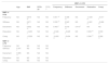
With the exception of the variables Decrement and Creep, PFM MMPs showed statistically significant differences between groups (p < 0.05). Both PFM tone and stiffness of the UUI group presented higher values and greater variability than the control group. However, the relaxation time was shorter in the group of women with UUI compared to the group of continent women. For the MMPs of LPM, only the Decrement showed no statistically significant differences between groups (p > 0.05). Women with UUI had less tone and stiffness, and higher values for VPs compared to women without UUI (Table 2).
Between-groups differences of muscle mechanical properties (MMP) of pelvic floor muscle (PFM) and lumbar paravertebral muscles (LPM).
| PFM | LPM | ||||||
|---|---|---|---|---|---|---|---|
| MMP | Control Group | UUI Group | P | Control Group | UUI Group | p | |
| Frequency (Hz) | 15.05 ± 1.97 | 16.26 ± 2.21 | 0.021 † | 17.10 ± 5.29 | 14.29 ± 3.44 | 0.012 † | |
| Stiffness (N/m) | 222.85 ± 62.67 | 259.18 ± 69.63 | 0.027 † | 356.41 ± 191.04 | 265.88 ± 109.81 | 0.020 † | |
| Decrement (Ø) | 1.04 ± 0.13 | 1.10 ± 0.16 | 0.080 | 1.28 ± 0.33 | 1.29 ± 0.35 | 0.872 | |
| Relaxation (ms) | 18.65 ± 2.94 | 17.03 ± 2.44 | 0.016 † | 17.42 ± 6.43 | 21.09 ± 5.67 | 0.015 † | |
| Creep (De) | 1.02 ± 0.13 | 0.97 ± 0.12 | 0.109 | 1.05 ± 0.35 | 1.25 ± 0.33 | 0.019 † | |
Values expressed as means ± SD.
For the within-group correlation analyses for the UUI group, the BMI showed a definite pattern, which was solely related to all MMPs of the PFM. This was a positive relation from moderate to strong with frequency, stiffness, and decrement, and negatively and fair to moderate with VP; in contrast, age only showed a positive correlation with stiffness and decrement of LPM. In addition, all MMPs of PFM were moderately related with at least three MMPs of LPM (|0.403|<r<|0.600|), with tone and stiffness of LPM showing the highest correlation values. Finally, none of the PF status questionnaires showed correlations with lumbopelvic MMPs (Table 3).
Correlations between muscle mechanical properties (MMP) of pelvic floor muscles (PFM) and lumbar paravertebral muscles (LPM), and age and body mass index (BMI), and pelvic floor questionnaires, for women with urge urinary incontinence (UUI).
| MMP of LPM | |||||||||
|---|---|---|---|---|---|---|---|---|---|
| Age | BMI | PFDI-20 | PFIQ-7 | Frequency | Stiffness | Decrement | Relaxation | Creep | |
| MMP of PFM | |||||||||
| Frequency | NS | 0.675 †† | NS | NS | 0.501 †† | 0.498 †† | NS | −0.445 †† | −0.417 † |
| Stiffness | NS | 0.671 †† | NS | NS | 0.532 †† | 0.543 †† | NS | −0.481 †† | −0.466 †† |
| Decrement | NS | 0.852 †† | NS | NS | 0.403 † | 0.518 †† | 0.415 † | NS | NS |
| Relaxation | NS | −0.493 † | NS | NS | −0.546 †† | −0.429 † | NS | 0.506 †† | 0.530 †† |
| Creep | NS | −0.359 † | NS | NS | −0.600 †† | −0.404 † | NS | 0.546 †† | 0.566 †† |
| MMP of LPM | |||||||||
| Frequency | NS | NS | NS | NS | |||||
| Stiffness | 0.341 † | NS | NS | NS | |||||
| Decrement | 0.695 †† | NS | NS | NS | |||||
| Relaxation | NS | NS | NS | NS | |||||
| Creep | NS | NS | NS | NS | |||||
Values expressed as Pearson's r or Spearman's rho coefficients.
In the control group, only age showed correlations, in this case with all LPM MMPs, and of moderate intensity: (frequency: r = 0.496, p = 0.003; stiffness: r = 0.506, p = 0.002; decrement: r = 0.402, p = 0.019; relaxation: r=−0.498, p = 0.003; creep: r=−0.425, p = 0.012). Similar to the UUI group, none of the questionnaires on pelvic floor status showed correlations with lumbopelvic MMPs. Finally, no correlation was identified between MMPs of PFM and MMPs of LPM.
DiscussionThe results showed differences in PFM and LPM MMPs according to the presence or absence of UUI. Women with UUI had higher tone and stiffness as well as shorter relaxation time in PFMs compared to continent women. However, the LPM of women with UUI had lower tone and stiffness, and higher VP compared to women without UUI. MMPs of women with UUI showed a homogeneous pattern of correlations, where PFM MMPs were associated with BMI, and with lumbar MMPs. Conversely, in healthy women, only age was moderately associated with LPM MMPs, and there was no relationship among lumbopelvic MMPs, either with each other or with BMI. Lumbopelvic MMPs were not related to the clinical status of the PF in either group.
Very limited information is available regarding the determination of MMPs in the PFM of women with UUI. Rodrigues-de-Souza et al22 observed good to excellent absolute measurement reliability for the determination of PFM tone and stiffness in women with and without UI. Our results showed mean between groups differences greater than the minimum detectable change for these properties.15,22 However, lower reliability values have been observed in the measurement of creep, as well as asymmetries between the sides of the perineum in some populations,14 which could explain the absence of differences between groups for this variable. In line with our results, previous studies have observed similar differences in PFM MMPs in other PFD,36,37 as well as an increase in tone accompanied by a decrease in the relaxation capacity of the PFM38 and motor control.37 In fact, women with provoked vestibulodynia have increased tone and stiffness, as well as decreased strength, speed of contraction, coordination, and endurance,39 which reinforces the existence of an altered pattern of MMP behavior of PFMs in different PFD, and specifically in UUI.
To our knowledge, this is the first study to analyze LPM MMPs in middle-aged women with UUI. However, the pattern of lower tone and stiffness, and higher VP values, observed in women with UUI in our study, has been similarly observed previously in vertebral algias.23 Along these lines, other authors have reported that muscle tone and stiffness is positively associated with the strength and thickness of lower extremity muscles40 and motor function of the upper extremities.41 Thus, the decrease in muscle tone and stiffness found could be explained by a reduction in the physical condition of women with UUI,42,43 related to the behavioral modifications observed in women with UI, where they may avoid certain activities for fear of urine leakage.43,44
The correlation between MMPs of PFMs and BMI observed in the women with UUI in our research concurs with the results reported by previous studies in different populations.14,45 This relationship is clinically relevant, as increased BMI is linked to the development of UUI.46 Similarly, the relationship between lumbopelvic MMPs in women with PFD found in our results supports the interdependence of both regions in the pathophysiology of UUI, as documented in low back pain.25,26 Finally, in line with our results, Alcaraz-Clariana et al19 found a positive correlation of age with increased tone, stiffness, and decreased LPM decrement in healthy women. This relationship was not observed in the MMPs of PFM, suggesting that other factors may be linked to the state of MMPs in the PF, at least in living women, because this relationship has been identified in cadavers.47,48
Regarding the clinical relevance of the tonometric analysis performed in the present study and its applicability, it is important to consider that UUI, as occurs with other PFD, is a very complex dysfunction due to the large number of underlying factors and mechanisms. Indeed, the PF can be thought of as a biomechanical structure due to the complex interaction between the vagina and its supportive structures that are designed to withstand the downward descent of the pelvic organs in response to increases in abdominal pressure.49 Therefore, a better understanding of UUI pathophysiology and the involvement of MMPs is relevant to advance treatments for this common disabling condition.7 There is a growing body of evidence to support the involvement of PFM tone and VP in the pathophysiology of specific PFD, such as vestibulodynia.36 In fact, the viscoelastic behaviour has an important functional significance for biological tissues. Tissues exhibiting a more pronounced creep behaviour, will stretch more under a constant load, and tissues presenting a higher relaxation behaviour will show a higher decrease in the stresses over time, when held at a constant length.50 In specific fields, such as childbirth, the knowledge of VP at the birth canal provides a better insight on how the duration of labour affects maternal adverse outcomes.51 Thus, the relaxation time property of the tissue contributes to reduce the damage levels in the second stage of labour, because this MMP allows the tissue to sustain less stiffness reduction, highlighting the importance of this feature in the maintenance of tissue integrity.52 Although animal models have been used to study elastic, relaxation time, and creep properties at the PF level,53,54 there is a need of instruments to obtain a better and more complete insight of MMPs assessment,36 such as tonometry. In summary, the present study obtained interesting results by jointly analyzing PFM and LPM MMPs in women with UUI, which, added to the innocuousness, speed, and low need for prior training of the tonometric protocol used, supports the study of MMPs in the clinical setting of physical therapy.36
Some limitations should be recognized. First, the external validity is limited to populations similar to those studied. In addition, the tonometric evaluation was always performed in a lying position, and different positions could lead to different results and interpretations.55 Moreover, the assessors were not blinded to the UUI status of the participants. Nonetheless, manual tonometry has low assessor dependence, which minimizes this potential source of bias.56 Further, longitudinal studies are required to analyze other potential factors that may modify lumbopelvic MMPs in women with UUI.
ConclusionThe study revealed that the presence of UUI may influence MMPs at PFM and LPM levels. Women with UUI have greater tone and stiffness as well as shorter relaxation time in PFM, whereas the LPM exhibited less tone and stiffness, and greater VP compared to women without UUI. The MMPs of women with UUI showed a homogeneous pattern of correlations, with MMPs of PFM being related with BMI, and with lumbar MMPs. The study highlights the clinical relevance of these associations and the need to consider both physical and clinical factors in managing UUI. These findings provide insights into the interconnected pathophysiology of UUI and other related musculoskeletal conditions. Finally, the determination of MMPs by externally applied hand-held instruments is clinically useful in the lumbopelvic region.
This research did not receive any specific grant from funding agencies in the public, commercial, or not-for-profit sectors.


