


Foot-ankle exercises targeting modifiable risk factors, such as peak pressure, ankle motion, and foot strength, may reduce ulcer risk and improve gait biomechanics, but the pathways driving these improvements are unclear.
ObjectivesTo identify the causal pathways through which a 12-week foot-ankle exercise program improved foot function, pain, and plantar pressure during gait by examining key mediators of these effects.
MethodsA total of 62 participants with diabetic peripheral neuropathy (DPN) were assigned to either a web-based foot-ankle program or usual care. The outcomes included peak pressure, pressure–time integral at the forefoot, foot pain, and foot function. Mediators included hallux and toe strength, sagittal ankle range of motion (ROM) during gait stance phase, ankle plantar flexion angle at push-off, ankle extensor moment at push-off, and foot pain and function. Outcomes and mediators were assessed at baseline, 12 weeks, and 24 weeks. Mediation models were tested using ordinary least squares regression with bias-corrected bootstrap confidence intervals.
ResultsThe analysis revealed consistent and inconsistent mediation effects. Improvements in foot function were mediated by reductions in foot pain (1.71, 95 % CI: 0.21, 4.43) and increases in ankle plantar flexion angle at push-off (0.45, 95 % CI: 0.15, 0.74). While the intervention directly reduced forefoot peak pressure, inconsistent mediation occurred, with indirect increases through reduced foot function (3.84, 95 % CI: 1.04, 11.38). Consistent mediation was observed, with increased sagittal ankle ROM during gait stance phase (9.02, 95 % CI: 2.47, 17.68) mediating a rise in the forefoot pressure-time integral.
ConclusionThe program reduced pain, improved function, and influenced plantar pressure through mediated pathways, highlighting a complex interplay of consistent and inconsistent mediation effects.
The literature highlights the need to understand the mechanisms through which complex physical therapy interventions produce their effects to ensure effective implementation and improve patient outcomes.1,2 The mediation analysis is one of the best choices for investigating these causal pathways, as it distinguishes between direct and indirect effects in randomized controlled trials.3 This statistical method identifies the pathways through which interventions have an impact, shedding light on the direct and indirect effects on outcomes.4 A secondary mediation analysis from resource-intensive trials could provide additional knowledge by identifying mediators driving the effects of a certain treatment and, thus, optimizing therapeutic interventions to target more effective clinical and functional outcomes critical to patients.
The international guidelines from the International Working Group on the Diabetic Foot (IWGDF) advise managing diabetic peripheral neuropathy (DPN) by addressing modifiable risk factors, such as abnormal plantar pressure, foot-ankle muscular weakness, and decreased foot-ankle mobility.5 Foot-ankle exercise programs have become a feasible conservative strategy to reduce ulcer risk by redistributing plantar pressure, increasing foot-ankle muscle strength, improving foot-ankle mobility, and optimizing gait biomechanics.6–8 The processes by which foot-ankle exercise therapies result in clinical, biomechanical, and functional changes are still unknown, despite growing evidence of their effectiveness. Understanding these pathways will provide crucial insights into the physiological mechanisms that drive DPN management as well as help to develop more targeted, evidence-based interventions.
Our research group has developed and validated a free, publicly available rehabilitation software, the Sistema de Orientação ao Pé Diabético – Diabetic Foot Guidance System (SOPeD),9 designed as an alternative to face-to-face physical therapy. The SOPeD targets musculoskeletal disorders associated with DPN. The SOPeD's effectiveness and cost-effectiveness were tested in the FOotCAre I (FOCA-I) trial, which demonstrated that at 12 weeks, the SOPeD web-based foot-ankle exercise program significantly improved foot function, reduced pain, enhanced ankle motion during gait, and optimized plantar pressure distribution in individuals with DPN.10 Additionally, SOPeD was shown to be a cost-effective approach compared to usual care, effectively reducing foot pain, DPN severity, and symptoms while also enhancing overall foot function.11 While these results emphasize the intervention's clinical benefits, the exact mechanisms behind these improvements remain to be elucidated. It is unclear if the improvements observed after the intervention were mediated by changes in foot muscle strength, foot function, ankle mobility, or other biomechanical or physical factors.
Therefore, this secondary mediation analysis of the FOCA-I study aims to clarify the causal pathways through which the SOPeD exercise program improved foot function, pain, and plantar pressure during gait by examining how these effects were mediated by key variables, breaking down the treatment effects into direct and indirect components.
MethodsStudy designThis study is a secondary data analysis from the FOCA-I randomized clinical trial that investigated the effects of the SOPeD web-based foot-ankle exercise program compared to usual care in treating modifiable risk factors for plantar ulcers. The detailed protocol and results of the trial are reported elsewhere.10,12 The trial was carried out at the Physical Therapy Department of the School of Medicine of the University of São Paulo, Brazil. This secondary analysis aimed to explore potential mechanisms underlying the treatment effect and was not prespecified in the protocol. All procedures were carried out in compliance with the Declaration of Helsinki, and informed consent was obtained from all study participants. The original study protocol was approved by the Ethics Committee of the School of Medicine at the University of São Paulo (CAAE: 90,331,718.4.0000.0065) and was registered at ClinicalTrials.gov on July 8, 2019 (NCT04011267). The trial's reporting adhered to the CONSORT guidelines,13 while the secondary analysis followed the Guidelines for Reporting Mediation Analyses of Randomized Trials (AGReMA).14
Participants and recruitmentIn total, 62 eligible participants ages 18 to 65 and classified as IWGDF risk categories 1 (low risk with loss of protective sensation [LOPS] or peripheral artery disease [PAD]) or 2 (moderate risk with LOPS and PAD, LOPS and foot deformity, or PAD and foot deformity) were randomly assigned 1:1 using a computer-generated sequence. Participants were recruited between August 1, 2019, and February 1, 2022, through social media, the database of the Endocrinology Outpatient Clinic at the Hospital das Clínicas (School of Medicine, University of São Paulo), and a campaign by the Brazilian Diabetes Care Association. All participants had a clinical diagnosis of type 1 or 2 diabetes, confirmed DPN using fuzzy software (www.usp.br/labimph/fuzzy; score ≥2), and could walk independently for at least 10 m Exclusion criteria included foot amputation, active ulcers, prior or indicated surgery on the lower limbs, severe unrelated neurological diseases, dementia, ongoing physical therapy, use of offloading devices or walking aids, and significant vascular complications or severe retinopathy. Additionally, all participants were required to have digital literacy.
Treatment armsThe control group (CG) and intervention group (IG) participants received self-care education, a self-management consultation from the main researcher, and a personalized brochure containing summarized self-care instructions according to the IWGDF guidelines.5 Participants in both groups maintained their ongoing medical and pharmacological treatments as the healthcare team prescribed.
Participants in the IG accessed the web-based foot-ankle exercise program via SOPeD. This program included three weekly sessions over 12 weeks, totaling 36 sessions, each lasting between 20 and 30 min. The exercises focused on strengthening and stretching both extrinsic and intrinsic foot muscles, with intensity adjusted according to each participant's perceived effort as recorded in the software. To enhance adherence, SOPeD incorporated gamification features and encouraged participants to complete reassessments every 30 days to ensure compliance with the exercise program.
During the first in-person session, IG participants received guidance on using SOPeD and executing the exercises along with supporting materials. A physical therapist remotely supervised the following 35 sessions, checking in with participants weekly to monitor their progress and software usage. Participants were instructed to report any adverse events and to discontinue the program if they experienced pain, excessive fatigue, or discomfort. Continuous oversight was maintained throughout the 12-week program, and participants were encouraged to continue with their exercise routine during the follow-up (24 weeks).10,15
OutcomesPeak pressure and pressure–time integral were measured using the emed-q pressure platform (novel GmbH, Munich, Germany) at 100 Hz. Participants walked barefoot over the platform at a self-selected pace for six trials. Only the forefoot region was selected for this secondary analysis as it showed pressure changes due to the intervention. The average of the six trials for each side was used for analysis.16
Foot pain and foot function were evaluated using the Brazilian version of the Foot Health Status Questionnaire (FHSQ).17 Two domains were assessed: foot pain (based on pain frequency, intensity, and impact on daily activities) and foot function (based on difficulties performing foot-related tasks like walking or standing). Scores range from 0 to 100, with higher values indicating better foot condition.
MediatorsMediators included isometric strength of the hallux and toes measured in a standing position using the emed-q100 pressure platform following the protocol by Mickle et al.18 The maximum force (N) was normalized to body weight and analyzed separately for the hallux and toe areas using the standard mask from the novel multimask software v.9.35 (novel GmbH).
Sagittal ankle range of motion (ROM) during gait stance phase, ankle plantar flexion angle at push-off, as well as the ankle extensor moment at push-off, were measured using a motion capture system (Vicon VERO; Oxford Metrics, Oxford, UK with eight infrared cameras) and a force plate (AMTI OR-6–1000; AMTI, Watertown, MA, USA); the variables were calculated using Nexus 2.6 software (Oxford Metrics). A total of 42 passive reflective markers were placed on both lower limbs following the Plug-In Gait and Oxford Foot Model protocols.19 Participants walked along a 10-meter track at a self-selected pace, and the average of five trials for each side was used for analysis.16
Foot pain and function were not only outcomes but also considered mediators, as pain could influence function, and function could affect plantar pressure distribution during gait. Table 1 presents the selected mediators and the theoretical rationale for their inclusion.
Description of selected mediators and theoretical rationales.
| Selected mediators | Theoretical rationale |
|---|---|
| Foot muscle strength | Foot-ankle exercises are commonly associated with both strengthening the foot-ankle muscles and reducing foot pain, particularly in musculoskeletal conditions.6 While strengthening exercises have long been linked to pain reduction,6 when the potential mediating effect of muscle strength on pain reduction was specifically investigated by a previous study, the results indicated that strength did not mediate the reduction in pain.20 This suggests that, although foot-ankle exercises may contribute to improved muscle strength and pain reduction, the precise mechanism through which pain reduction occurs remains unclear. Therefore, it is essential to further explore whether strengthening the foot muscles truly acts as a mediator in reducing foot pain. |
| Ankle motion during gait | Foot-ankle exercises have been shown to increase sagittal ankle ROM during gait stance phase and the ankle plantar flexion angle at push-off, as well as plantar pressure distribution.10,7 However, it remains unclear whether these changes are directly attributable to the exercises or if the modifications in ankle ROM and plantar flexion at push-off could mediate the effect on plantar pressure redistribution. Therefore, this potential mediating effect should be thoroughly investigated. |
| Ankle extensor moment at push-off | A previous study observed that foot-ankle exercises altered joint moments and plantar pressure distribution during gait.7 However, it is still unclear whether the changes in both outcomes were directly due to the exercises themselves or if modifying the joint moments could have mediated the effect on plantar pressure redistribution. Specifically, while strengthening the extensor muscles may influence ankle biomechanics and result in changes to plantar pressure distribution, the precise relationship between these effects and their underlying mechanisms remains to be fully understood. |
| Foot function (Foot Health Status Questionnaire) | In our published randomized controlled trial evaluating the effectiveness of foot-ankle mobility exercises and strengthening, we observed both improvements in foot function and changes in plantar pressure distribution during gait.10 Therefore, we would like to better understand whether this relationship directly resulted from the exercises in both outcomes or if improving foot function could have mediated the effect on plantar pressure distribution during gait. While foot-ankle mobility and strengthening exercises may optimize foot function and, in theory, modify plantar pressure distribution, there is still insufficient evidence to support this effect fully. |
| Foot pain (Foot Health Status Questionnaire) | Improved foot function, as a result of foot-ankle strengthening and mobility, may be explained by mechanical factors such as gains in mobility and strength. Studies have shown that reducing pain in musculoskeletal conditions can lead to an improved function.21 However, it remains unclear whether pain reduction acts as a mediator in this process or if both pain and function improvements are direct effects of the exercises. Further investigation is needed to clarify the interplay between these factors. |
Baseline values of both the mediator and the outcome were used to calculate the mean difference effect value. No confounding was assumed in the relationships between the intervention and the mediator or between the intervention and the outcome due to the random allocation of participants.
Time points and endpointsAll variables, including both outcomes and mediators, were assessed at baseline, 12 weeks, and 24 weeks by an assessor blinded to group allocation. For this mediation analysis, mediator data from the 12-week follow-up were used, while outcomes were evaluated at the 24-week follow-up. The 12-week time point was chosen for mediators to ensure temporal precedence, as it is assumed that changes in the mediators occur before changes in the outcomes.
Statistical analysisThe primary trial was powered for 62 participants, as reported in the trial protocol paper13; however, no formal sample size was calculated for the mediation analysis. All data were analyzed using SPSS Statistics v.21.0 (IBM, Armonk, New York).22 The baseline characteristics of the participants were reported as means and standard deviations or as numbers and percentages. Multiple imputation was employed to address missing data; the imputation model included age, sex, body mass index (BMI), type of diabetes mellitus, time of diabetes onset, and all available baseline and follow-up effect measure values. The dataset of this study was nearly complete, with a total dropout rate of 18 % (n = 11) across the entire sample. This dropout rate was 19 % in the IG (n = 6) and 16 % in the CG (n = 5). This dropout rate was anticipated in our sample size calculation, which included an expected dropout rate of 20 %.
The mediation models were tested using the PROCESS macro for SPSS,4 which is a tool for conducting mediation analyses through ordinary least squares regression. PROCESS utilizes bias-corrected bootstrap confidence intervals for inference about indirect effects, effectively addressing vulnerabilities to irregular sampling distributions that are common in the least squares regression.4 Mediation models assessing the effects of the intervention on potential mediators, as well as the effects of the mediators on the outcomes, were tested using PROCESS Model 4.4 A confidence interval of 95 % and 5000 bias-corrected bootstrap samples were employed for all PROCESS tests. We evaluated the indirect effect of the mediators by multiplying Path A (the correlation between the independent variable and the mediator) and Path B (the correlation between the mediator and the dependent variable) to determine the statistical significance of each mediation model.4 The direct effect is the portion of an intervention's impact that cannot be explained by a mediator, whereas the indirect effect is the portion that is mediated.3 Two sensitivity analyses were performed. In the first, mediation models were repeated with the inclusion of potential confounding variables as covariates in the PROCESS model: age, sex, BMI, type of diabetes mellitus, and time of diabetes onset. In the second, multivariable mediation models were conducted to further explore and complement the findings obtained through the initially adopted univariable models.
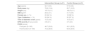
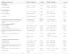
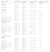
ResultsBaseline characteristics are shown in Table 2. The participant flow, follow-up assessment visit attendance, and reasons for dropout are detailed in a flowchart in Supplementary material online Figure S1. Table 3 presents Path A of the mediation analysis, which shows the estimated mean differences in the mediators, and Path B, which highlights the effect of the mediators on the clinical and biomechanical outcomes. Table 4 presents the indirect, direct, and total effects and the proportion of the effect mediated for all mediators and outcomes. Fig. 1 illustrates the significant mediating effects of mediators on the relationship between the randomized treatment groups (IG versus CG) and the outcomes.
Baseline clinical, demographic, and anthropometric characteristics for the intervention and control groups (n = 62).
Data are presented as mean ± standard deviation or as n ( %); Abbreviations: FHSQ, Foot Health Status Questionnaire; DPN, diabetic peripheral neuropathy; BMI, Body Mass Index.
Effect of the randomized treatment group on potential mediators (path A) and the effect of the mediators on the outcomes (path B), from the linear regression models.
Data are presented as mean (95 % confidence interval). ROM: range of motion.
Total, direct, and indirect effect estimates of randomized treatment group on the outcomes.
| Mediator/Outcome | Total Effect (Path C) | Direct Effect (Path C’) | Indirect Effect (Path A × Path B) | Proportion of the effect Mediateda |
|---|---|---|---|---|
| Foot pain | ||||
| Toes strength | 13.93 (6.65, 21.21) | 13.87 (6.57, 21.17) | 0.06 (−0.53, 0.68) | 0.4 % |
| Hallux strength | 13.40 (6.39, 20.40) | 14.04 (6.88, 21.20) | −0.64 (−2.07, 0.63) | 4.8 % |
| Foot function | ||||
| Toes strength | −0.75 (−7.65, 6.15) | −0.50 (−7.39, 6.38) | −0.24 (−1.54, 0.38) | 32.0 % |
| Hallux strength | −0.69 (−7.36, 5.97) | −1.97 (−8.77, 4.82) | 1.27 (−0.01, 2.91) | 184.1 % |
| Foot pain | −0.75 (−7.40, 5.89) | −2.47 (−9.20, 4.25) | 1.71 (0.21, 4.43) | 228.0 % |
| Ankle plantar flexion angle at push off | −1.05 (−8.01, 5.90) | −2.57 (−9.51, 4.37) | 1.51 (0.33, 3.23) | 143.8 % |
| Sagittal ankle ROM during gait stance phase | 1.65 (−6.02, 9.34) | 2.48 (−5.31, 10.28) | −0.82 (−2.45, 0.51) | 49.7 % |
| Forefoot peak pressure | ||||
| Toes strength | −9.49 (−62.16, 43.18) | −15.08 (−68.11, 37.94) | 5.59 (−1.90, 17.91) | 58.9 % |
| Hallux strength | −11.26 (−62.00, 39.48) | 0.85 (−50.61, 52.31) | −12.11 (−32.82, 5.60) | 107.6 % |
| Ankle plantar flexion angle at push off | −10.67 (−63.47, 42.12) | −10.75 (−63.61, 42.09) | 0.08 (−4.12, 3.60) | 0.7 % |
| Sagittal ankle ROM during gait stance phase | −10.44 (−62.36, 41.47) | −16.82 (−69.33, 35.68) | 6.37 (−4.52, 19.23) | 61.0 % |
| Ankle extensor moment at push-off | −10.44 (−62.36, 41.47) | −8.55 (−57.95, 40.84) | −1.88 (−20.00, 14.37) | 18.0 % |
| Foot function | −7.91 (−31.12, 15.29) | −11.75 (−35.08, 11.56) | 3.84 (1.04, 11.38) | 48.5 % |
| Forefoot pressure-time integral | ||||
| Toes strength | 11.70 (−6.04, 29.45) | 12.01 (−5.92, 29.94) | −0.30 (−3.25, 2.67) | 2.6 % |
| Hallux strength | 10.51 (−6.59, 27.61) | 8.57 (−8.88, 26.02) | 1.93 (−5.06, 9.85) | 18.4 % |
| Ankle plantar flexion angle at push off | 10.27 (−7.45, 28.01) | 10.24 (−7.50, 27.99) | 0.03 (−1.41, 1.59) | 0.3 % |
| Sagittal ankle ROM during gait stance phase | 9.89 (−7.57, 27.36) | 0.86 (−15.80, 17.53) | 9.02 (2.47, 17.68) | 91.2 % |
| Ankle extensor moment at push-off | 9.89 (−7.57, 27.36) | 10.56 (−5.94, 27.08) | −0.67 (−6.96, 5.18) | 6.8 % |
| Foot function | −10.55 (−20.08, −1.03) | −11.74 (−21.37, −2.11) | 1.18 (−0.91, 3.69) | 11.2 % |
Data are presented as mean (95 % confidence interval).
 and the outcomes. (a) Foot pain mediates the effect of the intervention on foot function, with pain changes influencing function. (b) Ankle plantarflexion angle at push-off mediates the intervention")
These models highlight the mediating roles of various factors in the relationship between the randomized treatment groups (intervention versus control) and the outcomes. (a) Foot pain mediates the effect of the intervention on foot function, with pain changes influencing function. (b) Ankle plantarflexion angle at push-off mediates the intervention's effect on foot function. (c) Foot function mediates the intervention's effect on forefoot peak pressure. (d) Sagittal ankle ROM during gait stance phase mediates the effect on the forefoot pressure–time integral. Path A represents the effect of the independent variable on the mediators, while Path B shows the effect of the mediators on the outcomes, together capturing the indirect effects. Path C’ reflects the direct effect of the treatment on the outcomes.
Mediation analyses demonstrated significant intervention effects on the mediator and significant effects of the mediator on the clinical outcomes across multiple variables. Specifically, the intervention had a significant effect on foot pain (Path A coefficient = 13.38, p < 0.001), which in turn significantly affected foot function (Path B coefficient = 0.12, p = 0.010). Similarly, the intervention significantly influenced ankle plantarflexion angle at push off (Path A coefficient = 13.38, p < 0.001), which subsequently significantly affected foot function (Path B coefficient = 0.12, p = 0.010). Furthermore, the intervention showed a significant effect on foot function (Path A coefficient = −8.97, p = 0.006), which was associated with a reduction in peak pressure at the forefoot (Path B coefficient = −0.42, p = 0.020). Lastly, the intervention significantly increased the sagittal ankle ROM during gait stance phase (Path A coefficient = 1.32, p = 0.003), which in turn had a notable effect on the pressure–time integral at the forefoot (Path B coefficient = 6.79, p < 0.001). There was no evidence of mediation by any of the other potential mediators considered (Table 1), as both Path A and Path B were not significant (no indirect effect).
Fig. 1 presents the results of the four models that illustrate the effects of the intervention on different outcomes, which were influenced by mediator outcomes. In Model A (Fig. 1a), foot pain acts as a mediator between the intervention and foot function. In Model B (Fig. 1b), ankle plantarflexion angle at push-off mediates the relationship between the intervention and foot function. In Model C (Fig. 1c), foot function mediates the relationship between the intervention and peak pressure at the forefoot. Finally, in Model D (Fig. 1d), sagittal ankle ROM during gait stance phase mediates the effect of the intervention on the pressure–time integral at the forefoot.
Total (Path C), direct (Path C’), and indirect (Path A × path B) effects of the intervention on outcomesThe intervention demonstrated significant direct and total effects on both foot pain and pressure–time integral at the forefoot, indicating a direct relationship between the independent variable (intervention) and the dependent variables (clinical and biomechanical outcomes). Evidence of mediation (indirect effect) was found for foot function through foot pain, foot function through ankle plantarflexion angle at push-off, peak pressure at the forefoot through foot function, and pressure-time integral at the forefoot through sagittal ankle ROM during gait stance phase. Two sensitivity analyses were conducted to evaluate the robustness of the findings. In the first sensitivity analysis, mediation models were repeated while including potential confounding factors such as age, sex, BMI, type of diabetes mellitus, and time of diabetes onset. Detailed results of this analysis are presented in Supplementary material online Tables S1 and S2. In the second sensitivity analysis, we performed multivariate mediation models to explore the results obtained in the univariable models. The multivariable analysis further validated the robustness of the findings, while highlighting additional nuances in the mediation effects. Detailed results of the multivariable mediation analysis can be found in Supplementary material online Tables S3 and S4.
Three indirect effects were positive, whereas the total effect was negative, indicative of inconsistent mediation (suppression). Specifically, a reduction in foot pain mediated an improvement in foot function (indirect effect), while the intervention itself led to a reduction in foot function (direct effect); an increase in ankle plantarflexion during push-off enhanced foot function (indirect effect), whereas the intervention itself directly led to a reduction in foot function (direct effect); and diminished foot function increased forefoot peak pressure (indirect effect), while the intervention itself led to a decrease in forefoot peak pressure (direct effect).
Conversely, one indirect effect exhibited consistent mediation, specifically the pressure–time integral at the forefoot mediated through ankle ROM. In this case, an increase in sagittal ankle ROM during gait stance phase resulted in a higher pressure–time integral, aligning with the positive direct effect. There were no other mediation effects (indirect effects) led by any of the other mediators considered.
DiscussionThis study examined how the SOPeD exercise program improved clinical and plantar pressure outcomes in individuals with DPN, elucidating the mechanisms behind these improvements. In general, the mediation analysis showcased that both foot pain and ankle plantarflexion angle during push-off mediated the intervention's effects on foot function. Additionally, foot function mediated the intervention's impact on forefoot peak pressure, while sagittal ankle ROM during gait stance phase mediated its effects on the forefoot pressure–time integral. No mediation effects were observed for any other potential mediators considered.
The intervention reduced foot function (direct effect), possibly due to transient effects like exercise-induced fatigue, altered biomechanics, or early adaptation during the intervention. However, improvements were driven by positive indirect effects from reduced foot pain and increased ankle plantarflexion at push-off, with mediated proportions of 228.0 % and 143.8 %, indicating inconsistent mediation. Despite the initial decline, long-term gains were achieved through these key mediators, highlighting their importance in achieving functional gains (24 weeks). The IG showed reduced forefoot peak pressure compared to the CG, suggesting that the SOPeD program effectively reduced plantar pressure independently of foot function changes (direct effect). However, the intervention decreased foot function and indirectly increased forefoot peak pressure. This direct reduction may have been influenced by unmeasured factors like postural adjustments, small joint movements, or motor control changes, directly reducing plantar pressure regardless of foot function. The indirect increase mediated by foot function might reflect participants’ initial inefficient foot use due to reduced function, altered foot rollover mechanics, and shifting pressure forward. Despite this, the total effect remained negative, indicating the intervention reduced peak pressure compared to the CG. The mediated proportion was 48.5 %, meaning that nearly half the effect occurred via indirect pathways. This highlights the intervention's complexity—positive indirect effects of foot function on pressure were offset by direct reductions, illustrating suppression in mediation, where direct and indirect effects interact to shape a nuanced total outcome. The scenarios above exemplify inconsistent mediation models, where a suppression effect occurs when the direct and mediated effects of an independent variable on a dependent variable have opposite signs.23,24 In contrast, consistent mediation models feature direct and mediated effects that share the same sign.25 In the present study, we observed consistent mediation where the intervention subtly increased the forefoot pressure–time integral compared to the CG, independent of changes in sagittal ankle ROM during gait stance phase (with a small and non-significant direct effect). However, the indirect effect revealed that an increase in sagittal ankle ROM during gait stance phase was significantly associated with a rise in the forefoot pressure-time integral. These findings suggest that while the direct effect on the pressure–time integral was minimal, improvements in sagittal ankle ROM during gait stance phase played a significant role in increasing the forefoot pressure-time integral during gait. These findings suggest that while the direct effect on the pressure–time integral was minimal, 91.2 % of the impact on forefoot pressure–time integral was mediated by increased sagittal ankle ROM during gait, highlighting its key role in modifying plantar load and guiding pressure-optimizing interventions. Although the initial reduction in foot function led to a significant increase in forefoot plantar pressure during gait, with peak pressure rising from 586.7 kPa to 715.0 kPa, the total effect of the intervention reduced the peak pressure at the forefoot. Additionally, the increase in pressure–time integral from 199.9 kPa·s to 251.8 kPa·s, should not be interpreted as a negative implication. This finding may suggest an improvement in foot mechanics rather than a negative effect, as these pressure values still fall within the normal range observed in healthy individuals.26,27 Specifically, barefoot peak pressure typically averages around 750 kPa,26 while pressure–time integral values generally range between 100 and 300 kPa·s.27 A recent systematic review established that a peak pressure value of 750 kPa when barefoot walking serves as a safe threshold for minimizing the risk of ulceration in people with diabetes.28
In this study, participants exhibited lower-than-normal peak pressures and pressure–time integrals at baseline,26–28 likely due to the reduced sagittal ankle ROM during gait stance phase, which impaired effective foot rollover during gait. Restricted sagittal ankle ROM during gait stance phase can limit load transfer to the forefoot, resulting in abnormally low plantar pressures. The intervention improved the sagittal ankle ROM during gait, restoring physiological foot mechanics and more evenly pressure distribution. These changes reflect functional restoration toward a healthy gait pattern, not harm. We initially hypothesized that an improvement in foot strength could be a mediator of the reduction in the peak pressure and foot pain; however, our analyses showed that the small changes in foot strength did not significantly mediate the intervention's effects on reducing peak plantar pressure and foot pain. Interestingly, a study by Holden,20 who also conducted a secondary mediation analysis, hypothesized that strengthening the hip muscles might have mediated a reduction in patellofemoral pain, but this was not observed. Holden et al.20 found that kinesiophobia and catastrophizing were the mediators behind the observed decrease in pain. Thus, other psychological factors might have influenced the reduction in foot pain observed after the intervention.
A limitation of this exploratory mediation analysis is that it only partially explains outcome improvements, being constrained by original trial data. Unmeasured factors like muscle blood flow, psychological aspects, or neuromuscular adaptations may also have contributed and deserve future investigation. Our findings underscore the significant potential of targeted foot-ankle exercise programs as an evidence-based intervention to address both functional and biomechanical impairments in individuals with DPN. By mediating improvements in foot pain and ankle mechanics, this program offers a scientifically grounded approach to optimize patient outcomes. Understanding mediation effects reveals how foot-ankle exercises work, supporting more confident treatment planning. Suppression effects highlight the intervention's complexity. Integrating this program into DPN care may enhance ankle mobility, reduce foot pain, and improve function, offering a comprehensive approach to managing DPN chronic complications. Furthermore, this study contributes to the growing body of evidence supporting the use of foot-ankle exercises in the treatment of musculoskeletal complications in individuals with DPN, strengthening the recommendation by the IWGDF for their inclusion in clinical care.5,6
While the uncertainty in direct and total effects reflected the complexity and variability of the intervention effects, the significant indirect effects underscore the robustness of mediated pathways. Given the study design and DPN population, these findings are plausible and offer valuable clinical insights. However, generalizability may depend on similar populations and adherence to the exercise program.
To our knowledge, this is the first study to use mediation analysis to explore the mechanisms underlying changes led by a foot-ankle exercise intervention on clinical and plantar pressure outcomes in people with DPN. Future studies should include more physiological and psychological variables in trial planning and in the multiple regression models to better capture mediated effects and refine intervention strategies.
ConclusionThis study showcases the mechanisms underlying the effects of the SOPeD exercise program in reducing foot pain and enhancing ankle plantarflexion angle during push-off, which in turn mediated improvements in foot function in people with DPN. Additionally, we showed that an increase in the sagittal ankle ROM during gait stance phase mediated an increase in the pressure–time integral at the forefoot after 12 weeks of exercise. While foot strength did not mediate these effects, sagittal ankle ROM during gait stance phase played an important role in improving the plantar pressure distribution during barefoot gait, suggesting restored foot-ankle mechanics and improved foot rollover.
Authors contributionsAll authors have made substantial contributions to the manuscript, including its conception and design, data analysis and interpretation, drafting, and critical revisions for important intellectual content. They all approved the final version for submission. RHCJ was responsible for the study design, data analysis, and interpretation, as well as writing and submitting the manuscript. JSSPF contributed to the study design, data interpretation, and manuscript writing. ICNS was responsible for the study design, secured funding, interpreted the data, and was involved in writing and submitting the manuscript. All authors reviewed, provided feedback on, and approved the final manuscript. No professional writers were involved.
Data availabilityThe datasets generated and/or analyzed during the current study are accessible as anonymized data on the University's public repository at https://repositorio.usp.br/.
The authors declare that the research was conducted in the absence of any commercial or financial relationships that could be considered a potential conflict of interest.
The authors would like to thank Professor Ana Claudia Latronico Xavier and the Endocrinology department for supporting us with a database of patients with diabetes at Hospital das Clínicas, Faculty of Medicine, University of São Paulo. The authors are grateful to Alisson Amorim Siqueira for the help with the web-based software development and updates.
This work was supported by Sao Paulo Research Foundation [FAPESP 2021/00807-8, 2019/02522-0] and by the National Council for Scientific and Technological Development, Brazil (CNPq) [28/2018 FOCA Trial 407252/2018-5]. Sacco is supported as a fellow researcher in CNPq, Brazil (Process 302558/2022–5). The funders had no role in the design, execution, interpretation, or writing of the study and did not have any authority over any study activity or in the decision to submit the report for publication.


