Pelvic organ prolapse (POP) affects many women and impacts quality of life. Evidence on physical activity in this population is inconsistent, particularly across the postoperative period. Objective: To describe patterns of physical activity and evaluate their associations with symptoms and QOL before and after reconstructive prolapse surgery.
MethodsThis secondary analysis of a randomized clinical trial used a descriptive and exploratory approach. A total of 466 patients were divided into two groups based on prolapse stage: stage II (n = 137) and stage III-IV (n = 329). Physical activity was measured using the metabolic equivalent task (MET). Secondary outcomes included the Pelvic Floor Distress Inventory (PFDI) and the Pelvic Floor Impact Questionnaire (PFIQ). The mixed model was used to analyze the MET, POP symptoms, and QoL at baseline, 3 months, and 12 months after surgery.
ResultsRegardless of prolapse stage, most patients were inactive at baseline (54.5%vs. 58.7%) and at 3 months (55.3%vs. 54.6%). By 12 months post-surgery, more patients became active (58.4%vs. 59.4%). No significant difference in MET was observed between groups over time (95% CI: [-219.9; 65.5]; p = 0.29). Similarly, no significant differences were found in symptoms and QoL (p > 0.05 for all comparisons).
ConclusionIrrespective of prolapse stage, patients showed similar physical activity levels at baseline, 3- and 12-months after surgery. Although no inferences can be made regarding timing or effects of postoperative exercise, there were no differences in symptoms and QoL between the groups over the course of one year.
Pelvic organ prolapse (POP) refers to the descent of one or more pelvic organs, such as the bladder, uterus, or rectum, into or through the vaginal canal, resulting from weakened pelvic floor support.1 It is a common condition with prevalence estimates between 1% to 31% based on symptomatic assessments, 10% to 50% during pelvic examination, and 20% to 65% when both methods are employed.1,2 POP significantly reduces a woman's quality of life (QoL) and patients often report experiencing a vaginal bulge within the previous four weeks.3 Research suggests that women have a 12.6% lifetime risk of requiring surgical correction for POP by the age of 80.4 The primary objective of these surgeries is to restore anatomy and thus, improve QoL by effectively alleviating symptoms.5 It is estimated that aging population in the United States will result in 47% increase in pelvic floor restorative surgeries between 2010 and 2050 leading to more women exposed to these procedures and so it is important to evaluate their outcomes on these women.6
The etiology of POP is multifactorial. Factors such as obstetrical trauma, hysterectomy, age, physical exercise involving chronic or repetitive straining have been associated with its development.1,7 Many of these factors may contribute to POP due to changes in muscles, ligaments, and connective tissues.1 There are two opposing hypotheses regarding the effect of physical activity on the pelvic floor in elite athletes.8 One hypothesis suggests that regular physical activity could strengthen the pelvic floor muscles (PFM), leading to coordinated muscle contractions and morphological changes such as hypertrophy and shortening of surrounding muscles, potentially reducing the risk of pelvic floor dysfunction, including stress urinary incontinence (SUI), anal incontinence (AI), and POP.8 The opposing hypotheses postulates that increased intra-abdominal pressure (IAP) during physical activities, if not counteracted by strong or quick co-contraction of PFM, might stretch and weaken these muscles during exercise.8
Contradictory evidence exists in the literature on the effects of physical exercise on POP.9–13 Some authors indicate that physical activities such as household chores are not associated with POP,4,14–16 while others suggest the onset or worsening of POP symptoms is related to the intensity of the physical exercise.11,17 Given this discrepancy, it is important to understand whether level of physical activity may be associated with prolapse severity at baseline and how these levels relate to symptoms and quality of life before and after surgery. Therefore, the objective of this study was to describe patterns of physical activity and examine their association with prolapse severity, symptoms, and quality of life before and after reconstructive prolapse surgery.
Materials and methodsStudy populationThe data for the current study were extracted from the Outcomes Following Vaginal Prolapse Repair and Midurethral Sling (OPUS) trial conducted by the Pelvic Floor Disorders Network, a cooperative agreement network sponsored by the Eunice Kennedy Shriver National Institute of Child Health and Human Development (NICHD) .18 The OPUS was a multicenter, randomized, single-blind, sham-controlled surgical intervention trial. The data reported in this study were requested from the NICHD Data and Specimen Hub (DASH). Data sharing was approved following signing a data user agreement (DUA) with the NICHD.
The original study included 337 patients with vaginal bulge symptoms and anterior vaginal prolapse, assessed using the Pelvic Organ Prolapse Quantification (POP-Q) System, defined as point Aa at -1 cm (1 cm above the hymen). The control and experimental groups underwent vaginal apical and anterior prolapse repair. The experimental arm underwent a concomitant retropubic midurethral sling (tension-free vaginal tape sling; Ethicon, Cincinnati, OH), and the control arm underwent sham incisions.18,19 Women were excluded if they had undergone previous sling placement, were receiving treatment for stress urinary incontinence, had contraindications for a midurethral sling (prior urethral surgery or pelvic irradiation), were planning pregnancy in the first year after surgery, or had a history of two or more hospitalizations for medical illnesses in the previous year.
Enrollment for the study began in May 2007, and follow-up was completed in January 2011. The OPUS data files include 337 randomized participants and 129 from the non-randomized cohort, followed for 12 months post-surgery, measuring surgical success, adverse events, and QoL using validated questionnaires.18,19
We included all 466 patients and divided them according to the POP stage: stage II (n = 137), stage III (n = 290), and stage IV (n = 39) based on the POP-Q system. For the analysis, the groups were divided into two groups (i.e., stage II and stage III-IV). The inclusion criteria for the secondary analysis involved participation in the surgical intervention trial, while exclusion criteria included the absence of information on prolapse stage and baseline metabolic equivalent task (MET) data (before surgery) for the analysis of physical activity level.
OutcomesThe primary outcome of interest was the level of physical activity measured by the MET in minutes per week (min/week). A MET represents the ratio of the energy cost of a specific activity to the resting metabolic rate; 1 MET corresponds to the energy expended while sitting quietly.21 The calculation of MET in min/week is done by multiplying the MET value, as defined by the Compendium of Physical Activities, by the duration of the activity in minutes.21 Participants were classified as inactive and active. Active participants were further categorized into low, moderate, and high physical activity levels.20 Additional questions provided descriptive information on physical activities including 'How often did you perform physical activities that required a major effort', 'Days a week doing strenuous exercise (past month)', 'Minutes each day doing this exercise', and 'Frequency of major effort in physical activity (past month)'.
The secondary outcomes of this study were QoL and the impact and severity of prolapse-related symptoms, assessed using the Pelvic Floor Distress Inventory (PFDI)22 and the Pelvic Floor Impact Questionnaire (PFIQ) .22 The PFDI is composed of three questionnaires: the Urinary Distress Inventory (UDI), the Pelvic Organ Prolapse Distress Inventory (POPDI), and the Colorectal-Anal Distress Inventory (CRADI). The PFIQ consists of three questionnaires: the Urinary Impact Questionnaire (UIQ), the Pelvic Organ Prolapse Impact Questionnaire (POPIQ), and the Colorectal-Anal Impact Questionnaire (CRAIQ). Scores on the UDI and POPDI range from 0 to 300, and scores on the CRADI range from 0 to 400. Similarly, the UIQ, POPIQ, and the CRAIQ scores range from 0 to 400. Higher scores on both questionnaires represent worse symptoms and poorer QoL. Information on physical activity, symptoms, and QoL was available at baseline, 3 months, and 12 months.22
Demographic, clinical and other potentially confounding variable data were collected on case report forms during recruitment and randomization, at baseline visit prior to surgery. These confounders were selected based on evidence from the literature and their known relevance to the outcomes of interest. These included: age, race, marital status, education level, employment status, health insurance, and BMI. This secondary analysis was approved by the Institutional Review Board of our institution (protocol number 2023P001511).
Statistical analysisData were analyzed using SPSS version 28.0 (IBM Corp., Armonk, NY, USA), and the figures were constructed in GraphPad Prism version 9.0.0 (San Diego, CA, USA). Normality of variables was assessed using the Shapiro–Wilk test. The demographic and clinical data were presented as median and 25th-75th percentiles for continuous variables and as absolute and relative frequencies for categorical variables according to the POP stage. The chi-square test was applied to compare categorical variables (demographic and physical activity) between groups, and the Mann-Whitney and Kruskal-Wallis test were used to compare continuous variables (clinical, prolapse-related symptoms and QoL) between groups. For the comparison of time points (baseline, 3 months, and 12 months) within each group, the Friedman test was applied.
The mixed model was used to compare the variables (MET, UDI, POPDI, CRADI, UIQ, POIQ, and CRAIQ) between the groups (stage II and stage III-IV) over time (at baseline, 3 months, and 12 months after surgery). Prolapse stage, age, and time were included as fixed effects (with age treated as a covariate), and participants were modeled with random intercepts to account for individual variability. Missing values were imputed using the last observation carried forward (LOCF) method to preserve longitudinal comparisons across time points. The link function estimated the outcomes according to the distribution of the variables. All reported p-values for multiple comparisons have been adjusted using the Bonferroni method. In all analyses, a p-value of <0.05 was considered statistically significant.
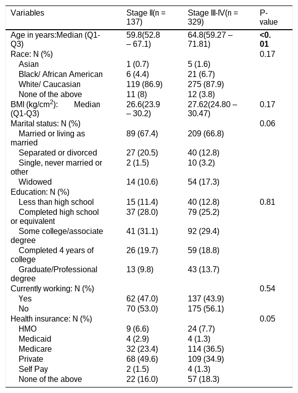
ResultsSociodemographic variablesA total of 466 patients with POP were included in this study. Table 1 represents the demographic characteristics according to the stage of POP. Those with stage III-IV POP were older on average (64.8 vs. 59.8; p < 0.01) as compared to stage II.
Sociodemographic variables by stage of pelvic organ prolapse at baseline.
Note: Q1 – 25th percentile. Q3 – 75th percentile. BMI – body mass index. The continuous variables are expressed as median and 25th and 75th percentiles and the categorical data are expressed as relative frequency (%) and absolute frequency (n). Identical letters indicate the difference between the times according to Bonferroni post-hoc. Bolded data indicate a significant difference (p < 0.05). Chi-square test applied to categorical variables and Kruskal-Wallis to continuous variables. Not all patients had a response recorded to every single question.
The majority of participants were white (86.9%; 87.9%), married or living as married (67.4%; 66.8%), and not currently working (53.0%; 56.1%). There was a similar distribution of race, BMI, marital status, health insurance and employment status among the three groups (Table 1).
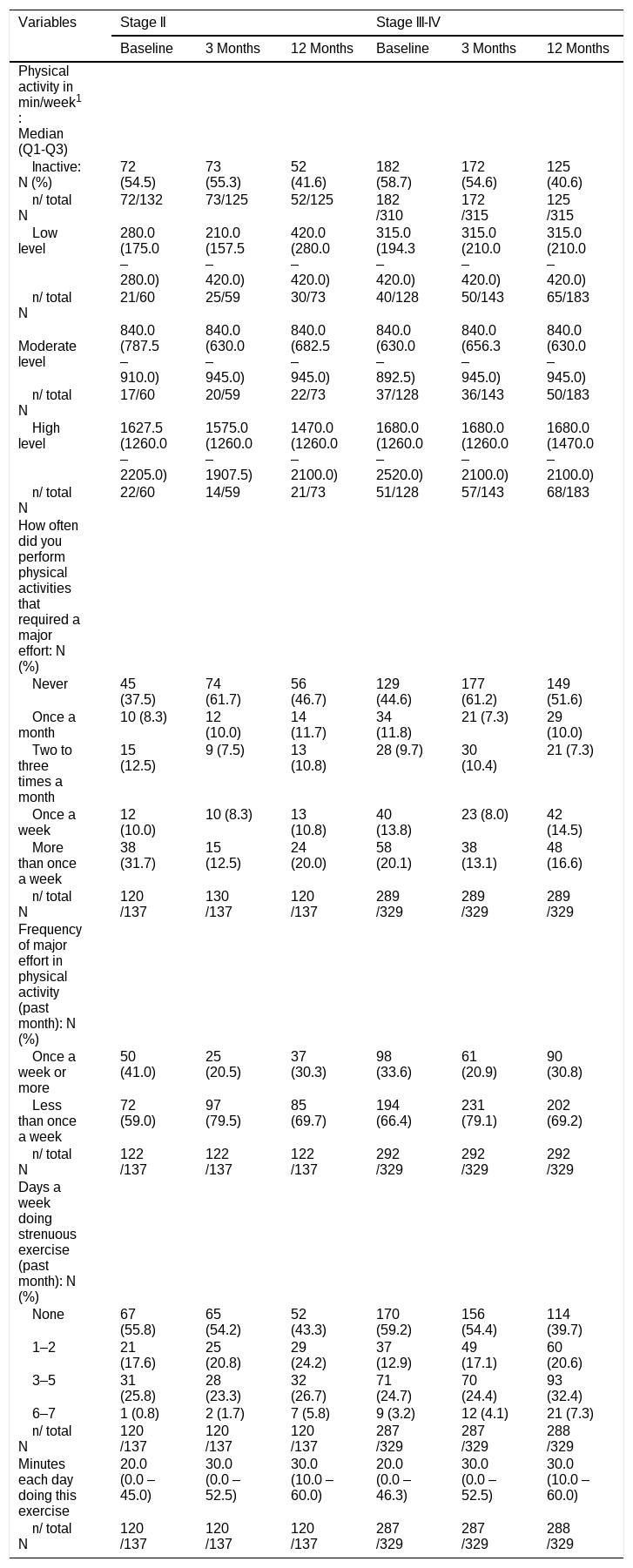
Physical activityTables 2 presents the level of physical activity in MET (min/week) of both groups at baseline, 3 months, and 12 months in addition to their response to questions related to physical activity practice. Regardless of the POP stage, most patients were inactive at baseline (54.5%vs. 58.7% p = 0.51) and at 3 months (55.3%vs. 54.6%; p = 0.91). However, by 12 months, a slightly higher number of patients were active in both groups (58.4%vs. 59.4%; p = 0.95). Baseline physical activity level was not associated with prolapse stage with similar distribution of low, moderate and high exercise level across the two groups (Table 3).
Profile of physical activity by stage of pelvic organ prolapse.
| Variables | Stage II | Stage III-IV | ||||
|---|---|---|---|---|---|---|
| Baseline | 3 Months | 12 Months | Baseline | 3 Months | 12 Months | |
| Physical activity in min/week1: Median (Q1-Q3) | ||||||
| Inactive: N (%) | 72 (54.5) | 73 (55.3) | 52 (41.6) | 182 (58.7) | 172 (54.6) | 125 (40.6) |
| n/ total N | 72/132 | 73/125 | 52/125 | 182/310 | 172/315 | 125/315 |
| Low level | 280.0(175.0 – 280.0) | 210.0(157.5 – 420.0) | 420.0(280.0 – 420.0) | 315.0(194.3 – 420.0) | 315.0(210.0 – 420.0) | 315.0(210.0 – 420.0) |
| n/ total N | 21/60 | 25/59 | 30/73 | 40/128 | 50/143 | 65/183 |
| Moderate level | 840.0(787.5 – 910.0) | 840.0(630.0 – 945.0) | 840.0(682.5 – 945.0) | 840.0(630.0 – 892.5) | 840.0(656.3 – 945.0) | 840.0(630.0 – 945.0) |
| n/ total N | 17/60 | 20/59 | 22/73 | 37/128 | 36/143 | 50/183 |
| High level | 1627.5(1260.0 – 2205.0) | 1575.0(1260.0 – 1907.5) | 1470.0(1260.0 – 2100.0) | 1680.0(1260.0 – 2520.0) | 1680.0(1260.0 – 2100.0) | 1680.0(1470.0 – 2100.0) |
| n/ total N | 22/60 | 14/59 | 21/73 | 51/128 | 57/143 | 68/183 |
| How often did you perform physical activities that required a major effort: N (%) | ||||||
| Never | 45 (37.5) | 74 (61.7) | 56 (46.7) | 129 (44.6) | 177 (61.2) | 149 (51.6) |
| Once a month | 10 (8.3) | 12 (10.0) | 14 (11.7) | 34 (11.8) | 21 (7.3) | 29 (10.0) |
| Two to three times a month | 15 (12.5) | 9 (7.5) | 13 (10.8) | 28 (9.7) | 30 (10.4) | 21 (7.3) |
| Once a week | 12 (10.0) | 10 (8.3) | 13 (10.8) | 40 (13.8) | 23 (8.0) | 42 (14.5) |
| More than once a week | 38 (31.7) | 15 (12.5) | 24 (20.0) | 58 (20.1) | 38 (13.1) | 48 (16.6) |
| n/ total N | 120/137 | 130/137 | 120/137 | 289/329 | 289/329 | 289/329 |
| Frequency of major effort in physical activity (past month): N (%) | ||||||
| Once a week or more | 50 (41.0) | 25 (20.5) | 37 (30.3) | 98 (33.6) | 61 (20.9) | 90 (30.8) |
| Less than once a week | 72 (59.0) | 97 (79.5) | 85 (69.7) | 194 (66.4) | 231 (79.1) | 202 (69.2) |
| n/ total N | 122/137 | 122/137 | 122/137 | 292/329 | 292/329 | 292/329 |
| Days a week doing strenuous exercise (past month): N (%) | ||||||
| None | 67 (55.8) | 65 (54.2) | 52 (43.3) | 170 (59.2) | 156 (54.4) | 114 (39.7) |
| 1–2 | 21 (17.6) | 25 (20.8) | 29 (24.2) | 37 (12.9) | 49 (17.1) | 60 (20.6) |
| 3–5 | 31 (25.8) | 28 (23.3) | 32 (26.7) | 71 (24.7) | 70 (24.4) | 93 (32.4) |
| 6–7 | 1 (0.8) | 2 (1.7) | 7 (5.8) | 9 (3.2) | 12 (4.1) | 21 (7.3) |
| n/ total N | 120/137 | 120/137 | 120/137 | 287/329 | 287/329 | 288/329 |
| Minutes each day doing this exercise | 20.0(0.0 – 45.0) | 30.0(0.0 – 52.5) | 30.0(10.0 – 60.0) | 20.0(0.0 – 46.3) | 30.0(0.0 – 52.5) | 30.0(10.0 – 60.0) |
| n/ total N | 120/137 | 120/137 | 120/137 | 287/329 | 287/329 | 288/329 |
Note:.
not included the inactive according to the metabolic equivalent of task. min/week – minutes per week.
2 total number of physically active individuals. The continuous variables are expressed in median and 25th and 75th percentiles and the categorical data are expressed in absolute frequency (N) and relative frequency (%). Q1 – 25th percentile. Q3 – 75th percentile. Not all patients had a response recorded to every single question.
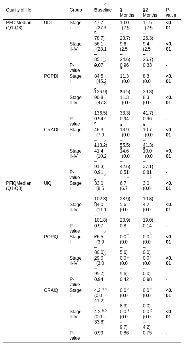
Comparison of quality of life by stage of pelvic organ prolapse at different time points.
Note: PFDI: Pelvic Floor Distress Inventory; UDI: Urinary Distress Inventory; POPDI: Pelvic Organ Prolapse Distress Inventory CRADI: Colorectal-Anal Distress Inventory; PFIQ: Pelvic Floor Impact Questionnaire; UIQ: Urinary Impact Questionnaire; POPIQ: Pelvic Organ Prolapse Impact Questionnaire (POPIQ); CRAIQ: Colorectal-Anal Impact Questionnaire. Q1 – 25th percentile. Q3 – 75th percentile. Superscript identical letters (a or b) the difference between the times according to Bonferroni post-hoc. Bolded data indicate a significant difference (p < 0.05). The continuous variables are expressed in median and 25th and 75th percentiles. Friedman test applied to compare the time (baseline, 3 months, and 12 months), and Mann-Whitney test to compare the groups (stage II and stage III-IV). Not all patients had a response recorded to every single question.
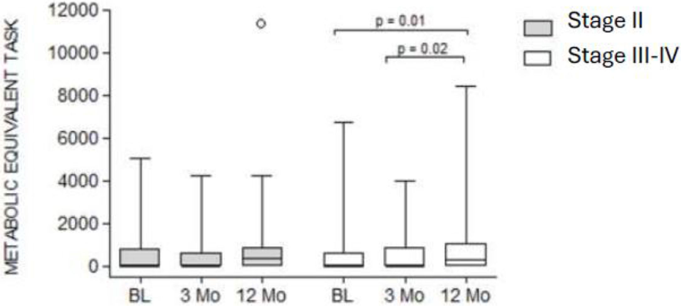
The mixed model showed a statistically significant difference in MET for patients with stage III-IV prolapse. This increase was observed between 3 and 12 months postoperatively (Δ = 280; 95% CI: –310.5 to –72.9; p = 0.02) and between baseline and 12 months (Δ = 280; 95% CI: –326.5 to –88.9; p = 0.01); positive Δ values indicate an increase in MET compared to the earlier time point. Conversely, no changes in MET were observed over one year in the stage II group. In addition, no significant interactions were detected, indicating that temporal trends did not differ between the groups (95% CI: –219.9 to 65.5; p = 0.29) (Fig. 1).

Comparison of the metabolis equivalent task across both groups over ane year.
A significant difference was observed over the course of one year for all QoL parameters, including at baseline, 3, and 12 months, when comparing data within each group individually. However, no significant differences were observed between the two groups (stage II and stage III-IV) at any time point (baseline, 3 months, and 12 months) (Table 3).
Mixed model analyses revealed no significant group-by-time differences for any of the pelvic floor symptom or quality of life measures. The coefficients for UDI (β = –492.8; p = 0.11), POPDI (β = –20.8; p = 0.06), CRADI (β = 172.0; p = 0.73), UIQ (β = –254.9; p = 0.71), POPIQ (β = –424.2; p = 0.52), and CRAIQ (β = –245.6; p = 0.72) all presented wide confidence intervals crossing zero, suggesting no association between POP stage and changes in symptom burden or quality of life over time.
DiscussionIn line with our aim to describe patterns of physical activity and evaluate their associations with symptoms and QoL before and after reconstructive prolapse surgery, our study showed that comparison between patients who underwent corrective surgery for stage II versus stage III-IV had similar levels of physical activity at baseline and throughout the year after surgery. Prolapse-related symptoms and QoL improved over time at 3 months and 12 months postoperatively. Additionally, we found a significant increase in MET for patients in the stage III-IV between the 3rd and 12th months, as well as between baseline and the 12th month.
Although women with stage III–IV prolapse demonstrated more advanced anatomical findings, symptom severity at baseline did not significantly differ from those in stage II. This is consistent with evidence showing that patient-reported symptoms do not necessarily progress in a linear fashion with anatomical stage.23 Symptom burden may vary across compartments and is not always proportional to POP-Q measurements.23 Furthermore, it has been reported that severe prolapse may mask symptoms of stress urinary incontinence due to urethral kinking,24 illustrating the complex and non-linear relationship between anatomical stage and symptom perception.
Few studies have examined the relationship between physical exercise and POP, indicating no significant association,12,14–16 as well as no correlation with the severity of POP.25 Similarly, our data confirm these findings of no difference in exercise intensity between stage II and stage III-IV up to one-year post-surgery. However, some studies suggest there might be an association between POP and exercise.11,17 Nygaard et al. (2016) described methodological challenges in research linking physical exercise with POP, including confounding factors, unclear occupational and activity histories, the use of non-standardized measures for POP outcomes, and the exclusion of domestic activities, which constitute a significant portion of daily activities for many women.11
One-year post-surgery, the majority of study participants reported a frequency of major effort in physical activity of less than once a week, which is different from current recommendations. Additionally, most patients indicated that they did not engage in strenuous exercise on any day of the week. In one study, physically active women who engage in heavy weightlifting as part of their exercise routine do not show a higher prevalence of POP symptoms.26 A case-control study involving 1610 women found no evidence that lifetime physical activities, including strenuous activities performed throughout their lives, increase the likelihood of POP in middle-aged women, except for possibly very high levels of strenuous activity during adolescence.27
Despite significant improvements in prolapse-related symptoms and QoL, there was no difference between the groups one year postoperatively. These results likely reflect the beneficial effects of surgical correction of prolapse, independent of prolapse stage, while also suggesting improvements that are independent of the level of physical activity. It is known that regular physical exercise offers numerous health benefits including improved QoL.28 Although some strenuous activities may damage the pelvic floor, it has been shown that regular low-impact activity, like walking, is associated with a lower prevalence of pelvic floor disorders.13
The data from this study showed that most patients were inactive at baseline, regardless of the level of POP. Nygaard et al. (2008) suggested that many women may be sedentary due to the physical, mental, and psychological burden of POP.29 The fear of worsening POP by engaging in physical activities is one of the self-imposed restrictions by women.30 Discomfort during physical exercise is a common reason for seeking health services.31
In our study, three months postoperatively, most patients remained inactive regardless of the group. A clinical trial aimed to compare patient satisfaction and pelvic floor symptoms after prescribing two forms of postoperative activity recommendations – one liberal and the other more restrictive – following reconstructive prolapse surgery showed that satisfaction was higher three months after surgery for women who were instructed to resume activities liberally.32 It was also observed that women in this group reported fewer prolapse and urinary symptoms and had similar short-term anatomical outcomes. The authors suggested that allowing women to resume normal activities postoperatively may lead to improvements in pelvic floor health.32
Interestingly, >40% of patients with POP remained inactive one year after surgery, despite improvements in QoL and pelvic symptoms. A study with 301 patients undergoing abdominal sacrocolpopexy indicated that only one-third of the women increased their exercise intensity and were less likely to report perceived interference with physical activity due to POP or its treatment.29 The same authors emphasized that there is insufficient scientific evidence to suggest an adverse effect of physical activity one year after prolapse surgery.29
Although most women resume physical exercise within a year after surgery, it is essential to provide guidance for those who remain inactive. There is a prevailing belief that engaging in physical exercise may increase the risk of POP recurrence, even after corrective surgery. Patients are advised to avoid specific activities during the recovery period to ensure the repair is not compromised before it has adequately healed. However, many of these activity restrictions have traditionally been based on limited or no solid evidence.33 Research indicates that physical exercise does not hinder the healing process.33,34 Typically, collagen deposition in the healing process begins on the second day, reaching its peak synthesis around the fifth day.35
Many restricted activities after pelvic floor surgery do not necessarily increase intra-abdominal pressure more than the act of standing up from a chair.31 Recently, three randomized clinical trials compared expedited activity with standard activity restrictions after POP surgery concerning anatomical outcomes, symptoms, and patient satisfaction.32,36,37 The findings indicate that expedited postoperative activity does not lead to inferior outcomes compared to those with more restricted activities.36,37 These studies support the resumption of physical activities ad libitum after minimally invasive prolapse surgery.32,36
The American College of Sports Medicine encourages most adults to engage in moderate-intensity cardiorespiratory exercise of ≥150 min/week or vigorous-intensity of ≥75 min/week. A combination of moderate and vigorous-intensity exercise is also an option with the goal of achieving a total energy of >500–1000 MET min/week.28 Nygaard et al. (2008) reported that 47% of patients who underwent abdominal sacrocolpopexy maintained unchanged exercise intensity, 36% increased their preoperative exercise intensity, and 18% decreased it. A minority (16%) participated in strenuous activities both before and after surgery.29
Our findings have several practical implications. Postoperative counselling should reassure patients about resuming normal activities once initial healing is achieved. Current evidence, including randomized trials, suggests that expedited resumption of activity after minimally invasive prolapse surgery does not compromise anatomical or functional outcomes.32,36,37 Enhanced Recovery After Surgery (ERAS) programs may provide a framework for standardized advice regarding postoperative activity and gradual return to exercise, emphasizing individualized approaches based on patient tolerance and comorbidities.38 Additionally, structured pelvic floor muscle training may be considered for patients who remain inactive or experience persistent symptoms, in line with recommendations from clinical guidelines. Overall, clinicians should provide evidence-based guidance to encourage safe physical activity, addressing patient fears regarding POP recurrence and promoting long-term health benefits.
The strengths of this study include its comprehensive analysis of a large randomized clinical trial with well-characterized data collected at baseline, 3 months, and 12 months. The use of MET allowed for a detailed characterization of physical activity levels, including functional and occupational activities, as well as specific questions regarding strenuous activities. Physical activity was assessed using self-reported data and expressed in MET values. This approach, although widely used in epidemiological and clinical research, is subject to recall bias and social desirability bias associated with self-reported questionnaire data and MET calculations. However, self-reported questionnaires are considered feasible and appropriate for large clinical trials and have been previously applied in studies with women undergoing pelvic surgery. Future research should consider incorporating objective measures, such as accelerometry or wearable activity trackers, to improve the accuracy of physical activity assessment.
As this study represents a secondary analysis, the potential influence of unmeasured confounders should also be considered, including prior physical activity history, variations in surgical techniques, and comorbidities that may influence postoperative physical activity levels. Moreover, a limitation of the study is the lack of information on participants' compliance with the postoperative instructions, and no data were collected on the participants' history of strenuous activities prior to surgery or other clinical conditions (disabilities, comorbidities, among others) that may interfere with physical exercise practice. There is no information available to justify the number of inactive individuals after one year. Data on general health over the course of one year and information on contraindications for physical exercise were not recorded in the primary study. In this exploratory study, using large, longitudinal data we were able to observe trends in certain subgroups, that could potentially be observed in daily clinical practice as patients with more advanced stages of POP tend to refrain from any exercise in fear of progressing it further as well as social shame that is linked to this condition refraining from e.g. group exercises. Thus, we believe correction of POP may lead to increased physical activity in real life scenario.
ConclusionOur findings indicated that patients with stage II and stage III-IV had similar levels of physical activity at baseline, 3 months, and 12 months following corrective POP surgery. Additionally, there was no significant difference when comparing symptoms and QoL between the two groups over the course of one year. These results describe patterns of physical activity and postoperative outcomes that did not differ by prolapse severity; however, these findings should be interpreted within the limitations of our dataset. Future studies should investigate optimal strategies for the gradual and safe implementation of exercise programs, as well as monitor physiological responses to exercise in active populations. Finally, new studies are required to answer the question of possible recurrence of prolapse after 12 months.
Authorship contributionsMM: Data analysis, Manuscript writing/editing.
RMG: Manuscript writing/editing.
IG: Manuscript writing/editing.
VAM: Manuscript writing/editing.
Financial disclaimers/conflict of interest statementNone.
Ethics statementInstitutional Review Board (IRB) of Mass General Brigham in Boston, Massachusetts, United States (protocol number 2023P001511).
The authors declare no competing interest
The authors acknowledge Eunice Kennedy Shriver National Institute of Child Health and Human Development (NICHD) for providing the “Outcomes Following Vaginal Prolapse Repair and Midurethral Sling (OPUS) trial” data that was used for this research. We also acknowledge John Wei, MD, the principal investigator who conducted the original study from which the data were generated. The content of this article is the responsibility of the authors and does not necessarily represent the views of the investigators of OPUS or the NICHD.